


Feeding 9 Billion: Innovations in Agricultural Modeling
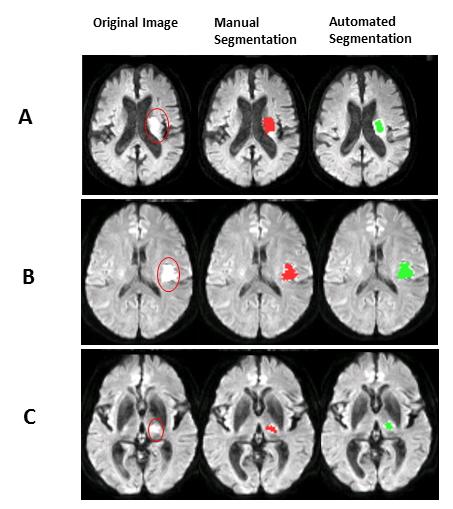
Examples of the manual and automated lesion segmentations. The first column shows the original DWIs, the second column shows the manual delineation of the acute ischemic lesions, and the third column demonstrate the results given by the proposed method.
Credit: Turku PET Centre
Lesion segmentation is a routine process where the abnormal areas within brain images are qualitatively and manually picked by expert radiologists.
However, manual lesion segmentation is time consuming and suffers from operator-bias. Accordingly, efficient and low-cost approaches for AIS lesion screening are yet to be introduced.
This research introduces a novel and fully automated technique for detection and segmentation of AIS lesions on MRIs and classification of images into stroke and none-stroke. This fully automated anomaly-detection method compares diffusion weighted images (DWIs) and apparent diffusion coefficients (ADC) images of the subjects with a group of healthy images in voxel-level.
Areas with hyperintensity on DWI and hypointensity on ADC are identified as lesions and saved as lesion masks. The lesion segmentation method was investigated on approximately 100 cases.
Since there is a risk of false lesion identification due to the artifacts, noises, and image low resolution, the lesion masks created by the method are screened and filtered via a binary classifier which either confirms that the created lesion mask contains a real AIS lesion or not. The classification performance was evaluated on about 200 MRIs.
The published results in the Journal of Neuroscience Methods show good agreement with the manually drawn lesions by experts (gold standard). The whole approach, including lesion segmentation and image classification, is straightforward, fast and does not require high computation power and memory.
“We believe that this method has the capacity to be implemented on an ordinary desktop workstation integrated into the routine clinical diagnostic pipelines of the hospitals.
This approach can help the radiologists to speed up the workflow of lesion detection and to reduce the operator bias in lesion segmentation owing to the reproducibility of the method”, tells project researcher Sanaz Nazari-Farsani from Turku PET Centre.


Synthetic biologists from Yale were able to re-write the genetic code of an organism — a novel genomically recoded organism (GRO) with one stop codon — using a cellular platform that they developed enabling the production of new classes of synthetic proteins. These synthetic proteins, researchers say, offer the promise of innumerable medical and industrial applications that can benefit society and human health. The creation of the landmark GRO, known as “Ochre” — which fully compresses redundant, or “degenerate” codons,…

Researchers work towards an inexpensive and portable solution for treating aphasia Electroencephalography (EEG) may offer a more accessible alternative to functional magnetic resonance imaging (fMRI) for guiding transcranial direct current stimulation (tDCS) when treating aphasia. Researchers from Institute of Science Tokyo found an 80% agreement between EEG and fMRI in identifying brain regions activated during language tasks. Furthermore, EEG-guided tDCS improved picture-naming speed in participants, indicating its potential for innovative therapies in language disorders. Many neurological disorders are directly linked…

Innovative stent surface technology to control vascular cell responses without drug side effects. The research team led by Dr. Hojeong Jeon and Dr. Hyung-Seop Han of the Biomaterials Research Center at the Korea Institute of Science and Technology (KIST, President Oh Sang-Rok), along with Dr. Indong Jun from KIST Europe, has developed a novel stent surface treatment technology using laser patterning. This technology promotes endothelial cell growth while inhibiting smooth muscle cell dedifferentiation in blood vessels. By controlling cellular responses to nanostructured…

Active monitoring is a sufficiently safe option when prostate MRI findings are negative. There are several strategies for the early detection of prostate cancer. The first step is often a blood test for prostate-specific antigen (PSA). If PSA levels exceed a certain threshold, the next step typically involves taking a tissue sample for analysis. Another option is to use magnetic resonance imaging (MRI) to search for signs of a tumor before deciding whether a biopsy is necessary, reserving biopsies only…


