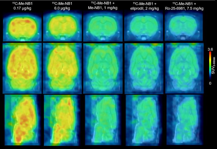

This figure shows rat brain 11C-Me-NB1 PET images (0-60 min) superimposed on an MRI template.
Credit: SD Krämer et al., ETH Zurich, Zurich, Switzerland
Swiss and German scientists developed the new PET radioligand, 11C-Me-NB1, for imaging GluN1/GluN2B-containing N-methyl-D-aspartate (NMDA) receptors (a class of glutamate receptor) in nerve cells.
When NMDA receptors are activated, there is an increase of calcium (Ca2+) in the cells, but Ca2+ levels that are too high can cause cell death. Medications that block NMDA receptors are therefore used for the treatment of a wide range of neurological conditions from depression, neuropathic pain and schizophrenia to ischemic stroke and diseases causing dementia.
“The significance of the work lies in the fact that we have for the first time developed a useful PET radioligand that can be applied to image the GluN2B receptor subunit of the NMDA receptor complex in humans,” explains Simon M. Ametamey, PhD, of the Institute of Pharmaceutical Sciences, ETH Zurich, in Switzerland.
“The availability of such a PET radioligand would not only help to better understand the role of NMDA receptors in the pathophysiology of the many brain diseases in which the NMDA receptor is implicated, but it would also help to select appropriate doses of clinically relevant GluN2B receptor candidate drugs. Administering the right dose of the drugs to patients will help minimize side-effects and lead to improvement in the efficacy of the drugs.”
For the study, 11C-Me-NB1 was used in live rats to investigate the dose and effectiveness of eliprodil, a drug that blocks the NMDA GluN2B receptor. PET scans with the new radioligand successfully showed that the receptors are fully occupied at neuroprotective doses of eliprodil. The new radioligand also provided imaging of receptor crosstalk between Sigma-1 receptors, which modulate calcium signaling, and GluN2B-containing NMDA receptors.
Ametamey points out, “These results mean that a new radiopharmaceutical tool is now available for studying brain disorders such as Alzheimer`s disease, Parkinson`s disease and multiple sclerosis, among others. It joins the list of existing PET radiopharmaceuticals used in imaging studies to investigate and understand underlying causes of these brain disorders.” He adds, “Furthermore, future imaging studies using this new radioligand would throw more light on the involvement of NMDA receptors, specifically the GluN2B receptors, in normal physiological processes such as learning and memory, as well as accelerate the development of GluN2B candidate drugs currently under development.”
###
Authors of “Evaluation of 11C-Me-NB1 as a potential PET radioligand for measuring GluN2B?containing NMDA receptors, drug occupancy and receptor crosstalk” include Stefanie D. Krämer, Thomas Betzel, Ahmed Haider, Adrienne Herde Müller, Anna K. Boninsegni, Claudia Keller, Roger Schibli and Simon M. Ametamey, Institute of Pharmaceutical Sciences, ETH Zurich, Zurich, Switzerland; Linjing Mu, University Hospital Zurich, Zurich, Switzerland; and Marina Szermerski and Bernhard Wünsch, University of Munster, Munster, Germany.
This study was financially supported by the Swiss National Science Foundation (Grant Number 160403).
Please visit the SNMMI Media Center to view the PDF of the study, including images, and more information about molecular imaging and personalized medicine. To schedule an interview with the researchers, please contact Laurie Callahan at (703) 652-6773 or lcallahan@snmmi.org. Current and past issues of The Journal of Nuclear Medicine can be found online at http://jnm.
ABOUT THE SOCIETY OF NUCLEAR MEDICINE AND MOLECULAR IMAGING
The Society of Nuclear Medicine and Molecular Imaging (SNMMI) is an international scientific and medical organization dedicated to advancing nuclear medicine and molecular imaging, vital elements of precision medicine that allow diagnosis and treatment to be tailored to individual patients in order to achieve the best possible outcomes.
SNMMI's more than 15,000 members set the standard for molecular imaging and nuclear medicine practice by creating guidelines, sharing information through journals and meetings and leading advocacy on key issues that affect molecular imaging and therapy research and practice. For more information, visit http://www.


