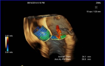

Valve geometry features are critical for disease diagnostics as well as surgical and catheter based therapy. Today physicians are performing valve measurement using 2D imaging only, making the decision process time consuming and operator dependent, which reduces its reproducibility.
Automated measurements are now enabled by a new ultrasound probe to reduce complexity that creates unstitched 3D images of the heart in real-time, combined with blood flow imaging via color Doppler technology.
From this image data, the eSie Valves advanced analysis package software enables efficient creation of a 3D model of the mitral and aortic valves, from which a multitude of measurements are computed. The eSie Valves package not only offers fast and reproducible quantification using clinical standard measurements, but also enables standard dynamic measurements of geometrically complex valve anatomy, which would not be practical to obtain manually.
eSie Valves is planned to be delivered with the new PRIME ACUSON SC2000 ultrasound systemPrime Ultrasound scanner.
This system offers a new trans-esophageal ultrasound probe. In practice, the transducer is inserted into the esophagus of a patient via an endoscope. In this way, the heart is imaged at close proximity, yielding highly accurate images. The device also measures the frequency of ultrasound waves reflected by blood cells (Doppler principle) and thereby computes the direction and speed of blood flow.
Learning software identifies heart valves automatically
In these images, eSie Valves automatically identifies heart valves and creates detailed 3D models. Image processing and machine learning technology, developed by Siemens' research division Corporate Technology, builds the foundation of the software. It enables fast and robust object detection within medical image data that is subject to noise and a wide spectrum of variation in appearance due to organ motion, pathology and patient variation. It is based on learning technology that analyzes hundreds of similar images from a database and learns how to identify recurrent image features as reference anatomical landmarks.
In the case of cardiac ultrasound images, a large number of acquisitions from different patients were used for the learning process. The software learns to identify certain anatomical features of different granularity, e.g. the coarse appearance of valves and chambers or fine details such as tips of the mitral valve. Then the software scans the image to determine location and pose of the valves, to finally generate a 3D model of the valve anatomy in a matter of seconds.
In collaboration with leading medical centers, clinical studies highlighted the reproducibility and speed of eSie Valves over competing solutions.
Weitere Informationen:
http://www.siemens.com/innovationnews


