


Feeding 9 Billion: Innovations in Agricultural Modeling
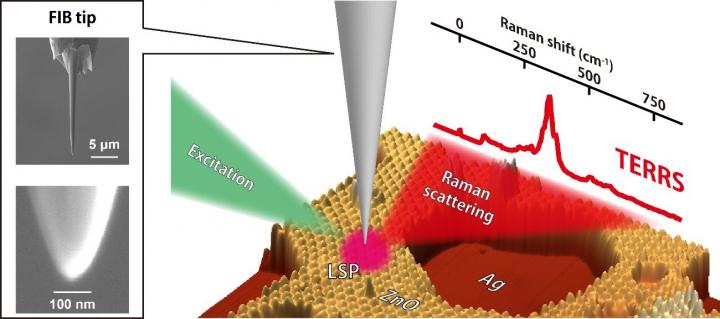
Tip-enhanced resonance Raman scattering is measured by a silver tip fabricated by focused ion beam (FIB) milling. Localized surface plasmon (LSP) is excited by an excitation laser, which generates enhanced Raman scattering from ultrathin zinc oxide (ZnO) films grown on a single-crystal silver (Ag) surface.
Credit: Takashi Kumagai
A research team at Fritz-Haber Institute in Berlin, headed by Dr. Takashi Kumagai, demonstrated tip-enhanced “resonance” Raman spectroscopy.
Resonance Raman spectroscopy is a powerful tool to analyze a specific chemical structure at a high sensitivity, but its spatial resolution has been restricted to be a few hundred nm due to the diffraction limit.
Extreme field confinement at a metal tip apex through localized surface plasmon excitation allows to break this limitation and now attain 1-nm resolution.
Tip-enhanced Raman spectroscopy takes advantage of atomic resolution imaging of scanning probe microscopy and enhanced Raman scattering through localized surface plasmon excitation.
The research team revealed tip-enhanced resonance Raman scattering in which both physical and chemical enhancement mechanisms are operative.
The underlying process was examined by modifying the localized surface plasmon resonance in the scanning tunneling microscope junction and by recording different-thickness zinc oxide films that exhibit a slightly different electronic structure.
In addition, the correlation between tip-enhanced resonance Raman scattering and local electronic states is resolved in combination with scanning tunneling spectroscopy that maps the local electronic state of the zinc oxide film.
The results explicitly show that a confined electromagnetic field can interact with local electronic resonances at the (sub)nanometer scale.


Discovery in a magnetic crystal could enable breakthroughs in quantum tech A team of Rice University researchers reported the first direct observation of a surprising quantum phenomenon predicted over half a century ago, opening pathways for revolutionary applications in quantum computing, communication and sensing. Known as a superradiant phase transition (SRPT), the phenomenon occurs when two groups of quantum particles begin to fluctuate in a coordinated, collective way without any external trigger, forming a new state of matter. The discovery…

‘Optical rotatum’ describes new structure of light Beams of light that can be guided into corkscrew-like shapes called optical vortices are used today in a range of applications. Pushing the limits of structured light, Harvard applied physicists in the John A. Paulson School of Engineering and Applied Sciences (SEAS) report a new type of optical vortex beam that not only twists as it travels but also changes in different parts at different rates to create unique patterns. The way the light behaves…

Although we know that supermassive black holes (millions of times the mass of our Sun) lurk at the centre of most galaxies, their very nature makes them difficult to spot and study. In contrast to the popular idea of black holes constantly ‘gobbling up’ matter, these gravitational monsters can spend long periods of time in a dormant, inactive phase. This was true of the black hole at the heart of SDSS1335+0728, a distant and unremarkable galaxy 300 million light-years away…

Despite its uniquely rich inventory of organic molecules, the moon may be able to support only a minuscule amount of biomass, a bioenergetic modeling study suggests. Titan, Saturn’s largest moon, is a strange, alien world. Covered in rivers and lakes of liquid methane, icy boulders and dunes of soot-like “sand,” its topography has long fascinated scientists and invited speculation on whether lifeforms might lurk beneath the moon’s thick, hazy atmosphere. An international team of researchers co-led by Antonin Affholder at…


