Medica 2019: Arteriosclerosis – new technologies help to find proper catheters and location of vasoconstriction
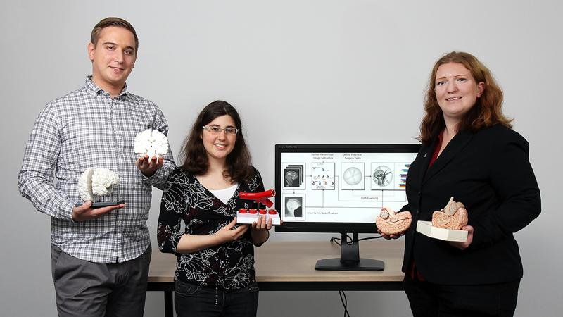
Robin Maack, Katharina Roth and Dr. Christina Gillmann (right) are developing these technologies.
Credit: Koziel/TUK
They will be presenting their technologies at the medical technology trade fair Medica held from 17 to 21 November in Düsseldorf at the Rhineland-Palatinate research stand (hall 7a, stand B06).
Catheters consist of different materials such as plastic, silicone or metal. These medical tubes are available in a number of variants, some are particularly easy to bend, others are rather rigid.
“Doctors often have to decide which catheters are best suited for treatment,” says computer scientist Robin Maack, who is working on the subject as part of his master's thesis at Technische Universität Kaiserslautern.
“It also plays a role, for example, whether the tube has to bend at a 90° angle in order to reach the affected area in the vessel,” continues Maack. The risk of injury needs to be as low as possible as well.
Together with Dr Christina Gillmann from the University of Leipzig, he is working on a database with which a suitable catheter can be found more quickly. For this purpose, they built a model for an artery that bends several times and branches at several points. The computer scientists record how long each catheter takes to reach the desired location and what forces act on the vessel wall to pass through the blood vessel.
“We analyse the videos and enter the results into our database,” says Gillmann. “For each model we provide important data such as material, size and manufacturer and then describe the most suitable areas of application.” In the future, physicians will be able to use the database to quickly find the right tube for treatment. This will also reduce the stress on patients in the future, as fewer catheters have to be tested in the body.
In addition, the researchers are working on a method that will show doctors exactly the location of the vascular constriction. “Blood vessels are usually very small, so that it is not always directly obvious where the constriction is,” explains the computer scientist. For this technology, images from Computed Tomography are used.
“With our analysis methods, we prepare the data of the images in a different way than is the case with conventional methods,” continues Maack. Moreover, the computer scientists subdivide the image data into the categories bones, veins, muscles and tissue types. Their computational program is able to detect even the smallest alterations in the image pixels and, in this way, to detect the affected area more reliably.
The researchers will present their work at the Medica in Düsseldorf.
Klaus Dosch, Department of Technology and Innovation, is organizing the presentation of the researchers of the TU Kaiserslautern at the fair. He is the contact partner for companies and, among other things, establishes contacts to science.
Contact: Klaus Dosch, Email: dosch[at]rti.uni-kl.de, Phone (also during the fair): +49 631 205-3001
Dr Christina Gillmann
University of Leipzig
Phone: +49 341 97-32373
E-mail: gillmann[at]informatik.uni-leipzig.de
Robert Maack
TU Kaiserslautern
Working Group Computer Graphics and Human Computer Interaction
Phone: +49 631 205-3268
E-mail: maack[at]rhrk.uni-kl.de


