Dead on against Tremors – Imaging technique improves tremor surgery

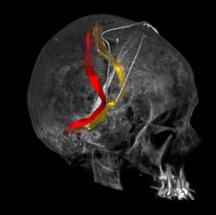
A patient's two-sided deep brain stimulation of the tremor bundles Medical Center - University of Freiburg/Volker Arnd Coenen
The idea of having a doctor operate on your open brain while you are wide awake does not sound pleasant. Unfortunately, however, deep brain stimulation in a fully conscious state is currently the only operative method available for many patients suffering from tremor diseases who can no longer tolerate the medications and long years of suffering brought on by their condition.
A Freiburg research team led by Prof. Dr. Volker Arnd Coenen, medical director of the Division of Stereotactic and Functional Neurosurgery at the Medical Center – University of Freiburg, has now succeeded in more accurately determining the position of the particular bundle of nerve fibers in the brain that deep brain stimulation needs to activate.
In the long term, the scientists hope this will allow surgeons to conduct deep brain stimulation to patients under general anesthesia, while at the same time reducing the risk of bleeding. The Freiburg researchers published the results of their study in the renowned journal Neurosurgery.
In their study on treating tremor-dominant Parkinson’s disease and essential tremor diseases by means of deep brain stimulation, the scientists compared the current method for detecting the fiber bundle with diffusion tensor tractography.
“This imaging technique produces images that are so precise that it is possible to determine the position of the fiber bundle in the brain with a margin of error of less than two millimeters. This reduces the amount of paths the electrodes need to take on the way to the target tissue in the brain, lowering the risk of vascular bleeding,” says Prof. Coenen.
Up to now, it was only possible to determine the target area indirectly on the basis of atlas data. The surgeons opens the skulls of fully conscious patients and conduct deep brain stimulation, steering the electrode toward regions in which they suspect that stimulation will lead to a reduction in the tremors.
If the point does not react to the stimulation, they remove the electrode and then direct it to another point via a new path from the surface. Each test increases the risk of damage to blood vessels and thus of vascular bleeding. The new method will make the operation safer for patients, as it will enable the surgeons to create a highly precise image of the tremor-reducing bundle structure directly.
The Freiburg researchers will soon begin two clinical studies applying this technology to essential tremor and Parkinson’s disease. The goal of the studies is to corroborate the findings from the current study.
Diffusion tensor tractography is an imaging technique that measures the diffusion of water molecules in body tissue with the help of magnetic resonance imaging (MRI) and represents it in spatially resolved form. It is particularly suitable for studying the brain because the diffusion behavior in the tissue undergoes characteristic changes in several diseases, allowing scientists to infer the course of the large bundles of nerve fibers. Diffusion tensor tractography has already been proven effective at detecting a new target site for stimulating the brain to treat depression, the medial forebrain bundle.
Deep brain stimulation influences and breaks up abnormal oscillations of nerve tissue with fine electric impulses. It requires the implantation of a brain pacemaker. The advantage of deep brain stimulation is that it provides constant, uninterrupted stimulation. When it is turned off, however, the symptoms return within minutes. Patients remain awake for most of the operation in which the neurostimuator is implanted, because “they help us to control the positioning of the electrodes,” says Prof. Coenen.
“We send a test impulse during the operation – when we’re at the right location, the patient’s symptoms, for instance trembling of the hands, are reduced immediately.” Currently, neurostimulation is not seen as a viable alternative until all other possible forms of therapy have been exhausted. But Prof. Coenen is confident: “Deep brain stimulation will gain importance as a therapy for various disorders.”
A tremor is defined as an involuntary, rhythmically repeating contraction of muscle groups that work in opposition to each other. The so-called physiological tremor can be measured, but it is almost impossible to see. A tremor only becomes visible when it appears as a symptom of a dis-ease, such as Parkinson’s disease.
The original publication, entitled “Modulation of the Cerebello-thalamo-cortical Network in Thalam-ic Deep Brain Stimulation for Tremor: A Diffusion Tensor Imaging Study,” is already available online and will also appear in the print version of Neurosurgery in December.
DOI: 10.1227/NEU0000000000000540
Contact:
Prof. Dr. Volker Arnd Coenen
Medical Director
Division of Stereotactic and Functional Neurosurgery
Phone: +49 (0)761 270-50630
volker.coenen@uniklinik-freiburg.de
Media Contact
More Information:
http://www.uniklinik-freiburg.deAll latest news from the category: Health and Medicine
This subject area encompasses research and studies in the field of human medicine.
Among the wide-ranging list of topics covered here are anesthesiology, anatomy, surgery, human genetics, hygiene and environmental medicine, internal medicine, neurology, pharmacology, physiology, urology and dental medicine.
Newest articles

Innovative 3D printed scaffolds offer new hope for bone healing
Researchers at the Institute for Bioengineering of Catalonia have developed novel 3D printed PLA-CaP scaffolds that promote blood vessel formation, ensuring better healing and regeneration of bone tissue. Bone is…

The surprising role of gut infection in Alzheimer’s disease
ASU- and Banner Alzheimer’s Institute-led study implicates link between a common virus and the disease, which travels from the gut to the brain and may be a target for antiviral…

Molecular gardening: New enzymes discovered for protein modification pruning
How deubiquitinases USP53 and USP54 cleave long polyubiquitin chains and how the former is linked to liver disease in children. Deubiquitinases (DUBs) are enzymes used by cells to trim protein…


