New High-Speed 3D Microscope—Scape—Gives Deeper View of Living Things

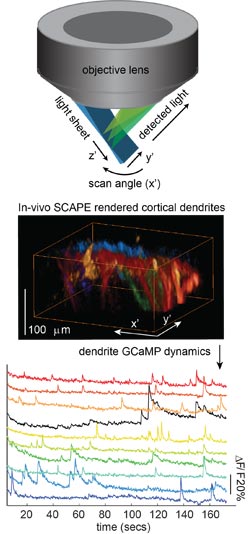
Elizabeth Hillman, Columbia Engineering SCAPE imaging geometry and neuronal firing in apical dendrites in mouse brain This schematic depicts SCAPE’s imaging geometry. The sample is illuminated by a thin sheet of light (blue), incident at an oblique angle. SCAPE achieves high speed imaging by sweeping this light sheet back and forth within the sample, achieved using a scanning mirror configured similarly to confocal microscopy. This optically sectioned plane is imaged onto a high speed sCMOS camera via the same objective lens. Unique de-scanning and image rotation optics ensure that the illuminated plane is always co-aligned with the camera plane, throughout its scan position. The end result is data equivalent to conventional light-sheet microscopy, but requiring a single, stationary objective lens, no sample translation, and consequently very high speed 3D imaging. This unique configuration permits volumetric imaging of intact tissues including the awake, behaving mouse brain. While limited in penetration depth (since SCAPE is currently implemented with a 488 nm laser) spontaneous activity in apical dendrites in layers 1 and 2 of the mouse cortex can be resolved at >10 volumes per second. Panels show dendrites rendered from SCAPE data acquired in an awake behaving mouse with layer 5 neurons labeled with GCaMP5g. Renderings show dendritic branches corresponding to the colored time-courses shown below. Temporal resolution and signal to noise are sufficient to discern different properties of onset and decay dynamics within individual dendritic branches for single events (see publication).
Opening new doors for biomedical and neuroscience research, Elizabeth Hillman, associate professor of biomedical engineering at Columbia Engineering and of radiology at Columbia University Medical Center (CUMC), has developed a new microscope that can image living things in 3D at very high speeds.
In doing so, she has overcome some of the major hurdles faced by existing technologies, delivering 10 to 100 times faster 3D imaging speeds than laser scanning confocal, two-photon, and light-sheet microscopy.
Hillman’s new approach uses a simple, single-objective imaging geometry that requires no sample mounting or translation, making it possible to image freely moving living samples. She calls the technique SCAPE, for swept confocally aligned planar excitation microscopy. Her study is published in the Advance Online Publication (AOP) on Nature Photonics's website on January 19, 2015.
“The ability to perform real-time 3D imaging at cellular resolution in behaving organisms is a new frontier for biomedical and neuroscience research,” says Hillman, who is also a member of Columbia’s Mortimer B. Zuckerman Mind Brain Behavior Institute. “With SCAPE, we can now image complex, living things, such as neurons firing in the rodent brain, crawling fruit fly larvae, and single cells in the zebrafish heart while the heart is actually beating spontaneously—this has not been possible until now.”
Highly aligned with the goals of President Obama’s BRAIN Initiative, SCAPE is a variation on light-sheet imaging, but, “It breaks all the rules,” says Hillman. While conventional light-sheet microscopes use two awkwardly positioned objective lenses, Hillman realized that she could use a single-objective lens, and then that she could sweep the light sheet to generate 3D images without moving the objective or the sample.
“This combination makes SCAPE both fast and very simple to use, as well as surprisingly inexpensive,” she explains. “We think it will be transformative in bringing the ability to capture high-speed 3D cellular activity to a wide range of living samples.”
SCAPE is an urgently needed breakthrough. The emergence of fluorescent proteins and transgenic techniques over the past 20 years has transformed biomedical research, even delivering neurons that flash as they fire in the living brain. Yet imaging techniques that can capture these dizzying dynamic processes have lagged behind. Although confocal and two-photon microscopy can image a single plane within a living sample, acquiring enough of these layers to form a 3D image at fast enough rates to capture events like neurons actually firing has become a frustrating road-block.
While SCAPE cannot yet compete with the penetration depth of conventional two-photon microscopy, Hillman and her collaborators have already used the system to observe firing in 3D neuronal dendritic trees in superficial layers of the mouse brain. In small organisms, including zebrafish larvae, SCAPE can see through the entire organism.
By tracking these tiny, unrestrained creatures in 3D at high speeds, SCAPE can capture both cellular structure and function and behavior. SCAPE can also be combined with optogenetics and other tissue manipulations during imaging because, unlike other systems, it does not require any movement of the imaging objective lens or the sample to create a 3D image.
Hillman and her students built their first SCAPE system using inexpensive off-the-shelf components. Her “aha” moment came when, looking at an old polygonal mirror in the lab, she realized how it could be used to generate SCAPE’s unusual scanning geometry. After several years of trial and error, Hillman and graduate student Matthew Bouchard came up with a configuration that worked, and beautiful images started to flow out. “It wasn’t until we built it that we realized it was a light-sheet microscope!” says Hillman. “It took us a while to realize how versatile the imaging geometry was, how simple and inexpensive the layout was—and just how many problems we had overcome.”
Beyond neuroscience, Hillman sees many future applications of SCAPE including imaging cellular replication, function, and motion in intact tissues, 3D cell cultures, and engineered tissue constructs, as well as imaging 3D dynamics in microfluidics, and flow-cell cytometry systems—all applications where molecular biology is delivering tools and techniques, but imaging methods have struggled to keep up. Hillman also plans to explore clinical applications of SCAPE such as video-rate 3D microendoscopy and intrasurgical imaging. Next-generation versions of SCAPE are in development that will deliver even better speed, resolution, sensitivity, and penetration depth.
As a member of the new Zuckerman Institute and the Kavli Institute for Brain Science at Columbia, Hillman is working with a wide range of collaborators, including Randy Bruno (associate professor of neuroscience, Department of Neuroscience), Richard Mann (Higgins Professor of Biochemistry and Molecular Biophysics, Department of Biochemistry & Molecular Biophysics), Wesley Grueber (associate professor of physiology and cellular biophysics and of neuroscience, Department of Physiology & Cell Biophysics), and Kimara Targoff (assistant professor of pediatrics, Department of Pediatrics), all of whom are starting to use the SCAPE system in their research.
“Deciphering the functions of brain and mind demands improved methods for visualizing, monitoring, and manipulating the activity of neural circuits in natural settings,” says Thomas M. Jessell, co-director of the Zuckerman Institute and Claire Tow Professor of Motor Neuron Disorders, the Department of Neuroscience and the Department of Biochemistry and Molecular Biophysics at Columbia. “Hillman’s sophistication in optical physics has led her to develop a new imaging technique that permits large-scale detection of neuronal firing in three-dimensional brain tissues. This methodological advance offers the potential to unlock the secrets of brain activity in ways barely imaginable a few years ago.”
Hillman’s technology is available for licensing from Columbia Technology Ventures and has already attracted interest from multiple companies.
This research was supported by the following grants: NIH (NINDS) R21NS053684, R01 NS076628 and R01NS063226, NSF CAREER 0954796, the Human Frontier Science Program and the Wallace H. Coulter Foundation (E.M.C.H.), NIH (NINDS) R01 NS069679 and the Dana Foundation (R.M.B.), (NINDS) R01NS070644 (R.S.M.), (NINDS) R01NS061908 (W.B.G.), DoD MURI W911NF-12–1-0594 (Yuste). M.B. received NSF and NDSEG graduate fellowships. V.V. was funded by an NSF IGERT Fellowship. C.S.M. is supported by a postdoctoral fellowship from Fundação para a Ciência e a Tecnologia, Portugal.
A patent related to this technique issued on December 31st 2013 (inventors Hillman and Bouchard). The authors are currently in licensing discussions.
Contact Information
Holly Evarts
Director of Strategic Communications and Media Rel
holly.evarts@columbia.edu
Phone: 212-854-3206
Mobile: 347-453-7408
Media Contact
All latest news from the category: Medical Engineering
The development of medical equipment, products and technical procedures is characterized by high research and development costs in a variety of fields related to the study of human medicine.
innovations-report provides informative and stimulating reports and articles on topics ranging from imaging processes, cell and tissue techniques, optical techniques, implants, orthopedic aids, clinical and medical office equipment, dialysis systems and x-ray/radiation monitoring devices to endoscopy, ultrasound, surgical techniques, and dental materials.
Newest articles

NASA: Mystery of life’s handedness deepens
The mystery of why life uses molecules with specific orientations has deepened with a NASA-funded discovery that RNA — a key molecule thought to have potentially held the instructions for…

What are the effects of historic lithium mining on water quality?
Study reveals low levels of common contaminants but high levels of other elements in waters associated with an abandoned lithium mine. Lithium ore and mining waste from a historic lithium…

Quantum-inspired design boosts efficiency of heat-to-electricity conversion
Rice engineers take unconventional route to improving thermophotovoltaic systems. Researchers at Rice University have found a new way to improve a key element of thermophotovoltaic (TPV) systems, which convert heat…


