Successful, Rapid Protein Crystallization Possible With Technique Developed by UCSD Researcher

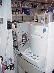
The mass spectrometer in the lab of Virgil Woods, M.D., constitutes a critical component of UCSD’s DXMS technology
An innovative method that allows increased success and speed of protein crystallization – a crucial step in the laborious, often unsuccessful process to determine the 3-dimensional structure unique to each of the body’s tens of thousands of folded proteins – has been developed by researchers at the University of California, San Diego (UCSD) School of Medicine and verified in tests with the Joint Center for Structural Genomics (JCSG) at The Scripps Research Institute (TSRI) and the Genomics Institute of the Novartis Research Foundation in La Jolla, California.
Described in the Jan. 20, 2004 issue of the journal Proceedings of the National Academy of Sciences (PNAS)*, the method, which employs a UCSD invention called enhanced amide hydrogen/deuterium-exchange mass spectrometry, or DXMS, rapidly identifies small regions within proteins that interfere with their ability to crystallize, or form a compact, folded state. The investigators demonstrate that once these regions are removed by what amounts to “molecular surgery”, the proteins then crystallize very well.
“Although the sequencing of the human genome gave us the code for genes that are the recipes for proteins, we need to see and understand the folded shape taken by proteins to determine how they work as the fundamental components of all living cells,” said UCSD’s Virgil Woods, Jr., M.D., the inventor of DXMS, senior author of the PNAS article and an associate professor of medicine. “Definition of a protein folded structure is of great use in the discovery of disease-targeting drugs. Furthermore, when we’re able to identify incorrectly folded proteins in disease states, such as Alzheimer’s, cystic fibrosis and many cancers, we may then be able to design drugs that encourage proper folding or block the misshapen protein.”
Unfortunately, many proteins do not naturally form a single, compact state in solution and hence, they are often highly resistant to crystallization, which is required for the x-ray crystallographic process that determines their shape. X-ray crystallography works by bombarding x-rays off crystals of a protein that contain a 3-dimensional lattice, or array of the individual protein or of a protein complex. The scattered, or diffracted pattern of the x-ray beams is used to calculate a s-dimensional structure of the protein.
“One of the major problems in trying to crystallize a protein is knowing whether it is really folded properly or has some disordered regions that might prevent crystallization,” said Ian Wilson, DPhil., JCSG principal investigator, one of the PNAS authors and a professor of molecular biology and Skaggs Institute for Chemical Biology at TSRI. “So, no matter how pure the protein might be, it might never crystallize without some further modifications to make it more compact and ordered.”
“We have used the information from DXMS to help guide our design efforts with protein targets that do not crystallize well,” said another of the paper’s authors, Scott Lesley, Ph.D., of the JCSG and the Genomics Institute of the Novartis Research Foundation. “Based on our experience to date, we have found that 20 to 40 percent of the targets that are amenable to crystallography could potentially benefit from the DXMS analysis. We think that DXMS analysis can be a key approach for salvaging problematic targets in structural genomics.”
In the experiments reported in PNAS, Woods and his team of researchers used the DXMS technology with 24 proteins provided by JCSG, a research consortium funded by the National Institute of General Medical Sciences (NIGMS), to generate 3-dimensional structures of proteins. Within a two-week period, DXMS provided data and analysis for 21 of the proteins sufficient to localize unstructured regions in the proteins, information that typically takes months, if not years, to obtain.
Recent studies have shown that many, if not the majority of proteins contain some unstructured, or unfolded regions of amino acid sequence interspersed with the structured regions. These unstructured regions appear to result in an inability of many proteins to crystallize.
Building upon previous research by Walter Englander, University of Pennsylvania, and David Smith, University of Nebraska, Woods has developed DXMS as a broadly applicable proteomics technology, and in the PNAS work, used it to rapidly and precisely identify the unstructured regions within proteins. The process measures the rate at which hydrogen molecules located within each amino acid of the protein, called peptide amide hydrogens, exchange with hydrogen in water in which the protein is dissolved. The rate of exchange depends on how exposed each amide hydrogen is to water in the folded protein. In unfolded regions of proteins, the amide hydrogens exchange at a much greater speed than do the amide hydrogens in the folded, structured portions of the protein. (For more information on DXMS, see Proceedings of the National Academy of Sciences 2003 June 10; 100(12):7057-62 http://www.pnas.org/cgi/content/full/100/12/7057 )
Of the 24 proteins provided by JCSG for DXMS analysis, six had already been crystallized and their structures determined. The results provided by DXMS matched the information on those six proteins, correctly identifying even small unfolded regions. The remaining 18 proteins provided by JCSG had all failed extensive prior crystallization attempts. In the new experiments, DXMS technology rapidly determined the unstructured regions in 15 of these proteins.
Two of the previously failed proteins were then subjected to “molecular surgery”, in which the DXMS-identified unstructured regions were selectively removed from the DNA that coded for the proteins. DXMS study of the resulting modified proteins demonstrated that the surgery had removed the unstructured regions without otherwise altering the shape of the originally well-folded regions. Each of the two resulting DXMS stabilized forms of the proteins were then found to crystallize well, while the original, unmodified proteins again failed to crystallize.
JCSG investigators were subsequently able to determine the 3-dimensional structures of these two proteins by x-ray analysis of the crystals resulting from DXMS-guided stabilization. One of the proteins that was successfully crystallized was found to have a unique shape or “fold”, not previously seen in proteins.
The research was funded by the National Institutes of Health Protein Structure Initiative Grant, the National Institute of General Medical Sciences, the University of California BIOSTAR and Life Sciences Informatics Program, and ExSAR Corporation.
Woods noted that the Innovative Molecular Analysis Technologies (IMAT) program of the National Cancer Institute has recently provided UCSD with a grant to further develop this application of DXMS. TSRI will be a subcontractor on that grant.
The University of California has patient applications pending on high-throughput DXMS and its application to crystallographic protein construct design as described in the PNAS paper.
Additional authors on the PNAS paper were Dennis Pantazatos, first author and a graduate student in the Biomedical Sciences Graduate Program of the UCSD School of Medicine; Jack S. Kim, UCSD Department of Medicine; Heath E. Klock, JCSG and the Genomics Institute of the Novartis Research Foundation; and Raymond C. Stevens, Ph.D., JCSG and TSRI.
* The PNAS paper, titled “Rapid refinement of crystallographic protein construct definition employing enhanced hydrogen/deuterium exchange MS,” was published Jan. 8, 2004 online, on the PNAS Early Edition http://www.pnas.org/cgi/reprint/0307204101v1
UCSD Contact:
Sue Pondrom
(619) 543-6163
spondrom@ucsd.edu
TSRI Contact:
Jason Socrates Bardi
858-784-9254
jasonb@scripps.edu
Media Contact
All latest news from the category: Life Sciences and Chemistry
Articles and reports from the Life Sciences and chemistry area deal with applied and basic research into modern biology, chemistry and human medicine.
Valuable information can be found on a range of life sciences fields including bacteriology, biochemistry, bionics, bioinformatics, biophysics, biotechnology, genetics, geobotany, human biology, marine biology, microbiology, molecular biology, cellular biology, zoology, bioinorganic chemistry, microchemistry and environmental chemistry.
Newest articles

Innovative 3D printed scaffolds offer new hope for bone healing
Researchers at the Institute for Bioengineering of Catalonia have developed novel 3D printed PLA-CaP scaffolds that promote blood vessel formation, ensuring better healing and regeneration of bone tissue. Bone is…

The surprising role of gut infection in Alzheimer’s disease
ASU- and Banner Alzheimer’s Institute-led study implicates link between a common virus and the disease, which travels from the gut to the brain and may be a target for antiviral…

Molecular gardening: New enzymes discovered for protein modification pruning
How deubiquitinases USP53 and USP54 cleave long polyubiquitin chains and how the former is linked to liver disease in children. Deubiquitinases (DUBs) are enzymes used by cells to trim protein…


