High-end microscopy reveals structure and function of crucial metabolic enzyme

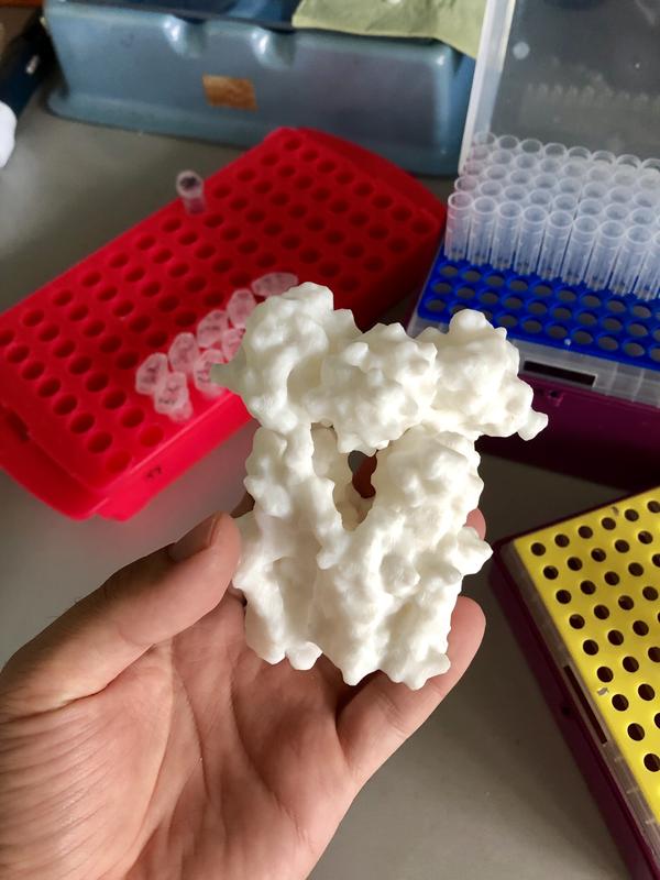
A 3D printed model of transhydrogenase. IST Austria – Domen Kampjut/Sazanov group
The enzyme transhydrogenase plays a central role in regulating metabolic processes in animals and humans alike. Malfunction can lead to serious disorders. For the first time, structural biologists at the Institute of Science and Technology Austria (IST Austria) have now visualized and analyzed the enzyme’s atomic structure with the support of the institute’s newly installed high-end cryo-electron microscope. The data presented in the journal Nature are relevant for the development of currently unavailable therapeutic options.
Within each cell, the power houses called mitochondria continuously break down molecules derived from food to generate energy as well as to produce new molecules that serve as building blocks of cells.
Balancing these two opposing processes is accomplished by an enzyme called proton-translocating transhydrogenase or NNT (nicotinamide nucleotide transhydrogenase). NNT sits in the mitochondria’s membrane and uses the electrochemical proton gradient generated by cellular respiration to provide the mitochondria with just the right amount of the co-enzyme NADPH, a vital metabolic precursor.
The proper functioning of NNT is crucial for metabolic regulation in all animals including humans. However, the details of how NNT accomplishes the coordinated transfer of protons across the membrane and synthesis of NADPH have remained obscure due to the lack of knowledge about the enzyme’s atomic structure.
Domen Kampjut, PhD student at IST Austria, and his supervisor and group leader Professor Leonid Sazanov have now for the first time visualized the molecule of mammalian NNT at a scale that allowed them to identify the structural principles of the enzyme’s channel gating—and thus to gain a deeper understanding of its functioning (and malfunctioning).
“Resolution revolution” at IST Austria
The atomic analysis of the enzyme NNT was only possible by taking advantage of state-of-the-art technology developments in cryo-electron microscopy (cryo-EM), the so-called “resolution revolution”. Parts of the current study’s data were generated using a new cryo-electron microscope installed at IST Austria only in fall 2018 and are the first to be published using one of three new machines, the “300 kV FEI Titan Krios”, in Klosterneuburg.
The cryo-EM analysis of NNT—which involved extensive time- and effort-consuming image processing and the support of the experts of the centrally organized and well-established Electron Microscopy Facility at IST Austria—delivered near atomic-resolution images of the molecule’s three different domains in various conformational states.
Opening the gate for protons—and new forms of medical treatment
With these images, the structural biologists could show how the domain that binds NADPH can open the proton channel to either side of the mitochondrial membrane. First author Domen Kampjut: “NNT has been studied for a few decades, but classical imaging methods such as X-ray crystallography have failed to give a detailed look into its structure because it is highly dynamic. Furthermore, membrane proteins like NNT are particularly challenging to study as they are fragile and difficult to purify in large amounts needed for crystallography. Thus, only with cryo-EM could we finally see clearly how the proton transfer works—and with this, find a missing piece of the puzzle on the way to understanding what to do if it does not work.”
Professor Leonid Sazanov adds: “These structures are particularly exciting because transhydrogenase performs an amazing volte-face by rotating an entire, quite large, NADPH-binding domain 180 degrees ‘up’ or ‘down’. This is, as far as we know, unique among studied enzyme mechanisms. However, such a rotation now makes complete sense in view of our proposed mechanism and it shows how nature can ‘creatively’ solve challenging tasks.”
The results are an important further step towards the development of novel therapies. For instance, the development of currently unavailable NNT inhibitors has great therapeutic potential with regard to metabolic dysfunctions including metabolic syndrome, and some cancers.
Background information
New cryo-EM at IST Austria—high-end technology to support high-end research
In 2018, IST Austria purchased and installed three state-of-the-art cryo-electron microscopes (cryo-EMs) as part of the established and centrally organized Electron Microscopy Facility of the institute, allowing scientists to observe biological structures at near-atomic scales. The technology of cryo-EM, which earned development contributors the 2017 Nobel Prize in chemistry, has led to a series of breakthrough discoveries in the life sciences in the recent years. Using cryo-EM, biological samples such as proteins can be observed in their natural state, rendering this method indispensable in structural biology.
IST Austria’s cryo-EM facility consists of one 300 kV, one 200 kV, and one cryo-dedicated focused ion beam scanning electron microscope (cryo FIB/SEM). The “300 kV FEI Titan Krios”, a cryo transmission electron microscope (cryo-TEM), used in this study is particularly noteworthy: “This machine is unique in Austria,” says facility manager Ludek Lovicar. “Currently, no other Austrian institution has a state-of-the-art 300 kV electron microscope under cryo conditions.” The current study demonstrates that scientific excellence paired with innovative technology can bring about groundbreaking research results and increase their application potential.
More information: https://ist.ac.at/en/research/scientific-service-units/electron-microscopy-facil…
Original publication:
Domen Kampjut & Leonid A. Sazanov. 2019. Structure and mechanism of mitochondrial proton-translocating transhydrogenase. Nature. DOI: 10.1038/s41586-019-1519-2
Grant information:
This project has received funding from the European Union’s Horizon 2020 research and innovation program under the Marie Skłodowska-Curie Grant Agreement No. 665385.
Originalpublikation:
https://www.nature.com/articles/s41586-019-1519-2
https://seafile.ist.ac.at/d/0441b17c26c7406cafe0/ Picture download
Media Contact
More Information:
https://ist.ac.at/de/All latest news from the category: Life Sciences and Chemistry
Articles and reports from the Life Sciences and chemistry area deal with applied and basic research into modern biology, chemistry and human medicine.
Valuable information can be found on a range of life sciences fields including bacteriology, biochemistry, bionics, bioinformatics, biophysics, biotechnology, genetics, geobotany, human biology, marine biology, microbiology, molecular biology, cellular biology, zoology, bioinorganic chemistry, microchemistry and environmental chemistry.
Newest articles

A ‘language’ for ML models to predict nanopore properties
A large number of 2D materials like graphene can have nanopores – small holes formed by missing atoms through which foreign substances can pass. The properties of these nanopores dictate many…

Clinically validated, wearable ultrasound patch
… for continuous blood pressure monitoring. A team of researchers at the University of California San Diego has developed a new and improved wearable ultrasound patch for continuous and noninvasive…

A new puzzle piece for string theory research
Dr. Ksenia Fedosova from the Cluster of Excellence Mathematics Münster, along with an international research team, has proven a conjecture in string theory that physicists had proposed regarding certain equations….


