New super-resolution technique allows for more detailed brain imaging

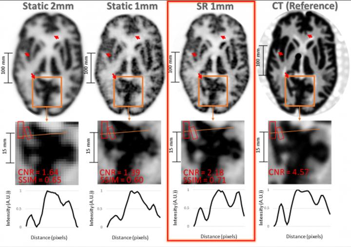
Result of the Hoffman brain phantom study. Top row: same PET slice reconstructed with A) 2mm static OSEM, B) 1mm static OSEM, C) proposed SR method and D) corresponding CT slice (note that the CT image can be treated as a high-resolution reference). Middle row: zoom on region of interest for corresponding images. Bottom row: Line profiles for corresponding data.
Image created by Y Chemli, et al., Gordon Center for Medical Imaging: Department of Radiology Massachusetts General Hospital, Harvard Medical School, Boston, MA.
A new imaging technique has the potential to detect neurological disorders–such as Alzheimer’s disease–at their earliest stages, enabling physicians to diagnose and treat patients more quickly. Termed super-resolution, the imaging methodology combines position emission tomography (PET) with an external motion tracking device to create highly detailed images of the brain. This research was presented at the Society of Nuclear Medicine and Molecular Imaging’s 2021 Virtual Annual Meeting.
In brain PET imaging, the quality of the images is often limited by unwanted movements of the patient during scanning. In this study, researchers utilized super-resolution to harness the typically undesired head motion of subjects to enhance the resolution in brain PET.
Moving phantom and non-human primate experiments were performed on a PET scanner in conjunction with an external motion tracking device that continuously measured head movement with extremely high precision. Static reference PET acquisitions were also performed without inducing movement. After data from the imaging devices were combined, researchers recovered PET images with noticeably higher resolution than that achieved in the static reference scans.
“This work shows that one can obtain PET images with a resolution that outperforms the scanner’s resolution by making use, counterintuitively perhaps, of usually undesired patient motion,” said Yanis Chemli, MSc, PhD, candidate at the Gordon Center for Medical Imaging in Boston, Massachusetts. “Our technique not only compensates for the negative effects of head motion on PET image quality, but it also leverages the increased sampling information associated with imaging of moving targets to enhance the effective PET resolution.”
While this super-resolution technique has only been tested in preclinical studies, researchers are currently working on extending it to human subjects. Looking to the future, Chemli noted the important impact that super-resolution may have on brain disorders, specifically Alzheimer’s disease. “Alzheimer’s disease is characterized by the presence of tangles composed of tau protein. These tangles start accumulating very early on in Alzheimer’s disease–sometimes decades before symptoms–in very small regions of the brain. The better we can image these small structures in the brain, the earlier we may be able to diagnose and, perhaps in the future, treat Alzheimer’s disease,” he noted.
Abstract 34. “Super-Resolution in Brain PET Using a Real Time Motion Capture System,” Yanis Chemli, LTCI, Telecom Paris, Institut Polytechnique de Paris, Paris, France, and Gordon Center for Medical Imaging, Boston, Massachusetts; Marc-Andre Tetrault, Computer Engineering, Universite de Sherbrooke, Sherbrooke, Quebec, Canada; Marc Normandin, Georges El Fakhri, Jinsong Ouyang and Yoann Petibonn, Department of Radiology, Massachusetts General Hospital, Harvard Medical School, Boston, Massachusetts; and Isabelle Bloch, Sorbonne Universite, CNRS, LIP6, Paris, France, and LTCI, Telecom Paris, Institut Polytechnique de Paris, Paris, France.
###
All 2021 SNMMI Annual Meeting abstracts can be found online at https:/
About the Society of Nuclear Medicine and Molecular Imaging
The Society of Nuclear Medicine and Molecular Imaging (SNMMI) is an international scientific and medical organization dedicated to advancing nuclear medicine and molecular imaging, vital elements of precision medicine that allow diagnosis and treatment to be tailored to individual patients in order to achieve the best possible outcomes.
SNMMI’s members set the standard for molecular imaging and nuclear medicine practice by creating guidelines, sharing information through journals and meetings and leading advocacy on key issues that affect molecular imaging and therapy research and practice. For more information, visit http://www.
All latest news from the category: Medical Engineering
The development of medical equipment, products and technical procedures is characterized by high research and development costs in a variety of fields related to the study of human medicine.
innovations-report provides informative and stimulating reports and articles on topics ranging from imaging processes, cell and tissue techniques, optical techniques, implants, orthopedic aids, clinical and medical office equipment, dialysis systems and x-ray/radiation monitoring devices to endoscopy, ultrasound, surgical techniques, and dental materials.
Newest articles

Innovative 3D printed scaffolds offer new hope for bone healing
Researchers at the Institute for Bioengineering of Catalonia have developed novel 3D printed PLA-CaP scaffolds that promote blood vessel formation, ensuring better healing and regeneration of bone tissue. Bone is…

The surprising role of gut infection in Alzheimer’s disease
ASU- and Banner Alzheimer’s Institute-led study implicates link between a common virus and the disease, which travels from the gut to the brain and may be a target for antiviral…

Molecular gardening: New enzymes discovered for protein modification pruning
How deubiquitinases USP53 and USP54 cleave long polyubiquitin chains and how the former is linked to liver disease in children. Deubiquitinases (DUBs) are enzymes used by cells to trim protein…


