3D liver aids surgery

The sharing of supercomputing resources via the Internet could allow vitual organs to become standard hospital decor
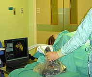
Virtual organ image beamed into OR.
Liver surgeon Rory McCloy carries a wad of CAT scans into his operations. “I spend my life looking at 60 slices of salami,” he says. Peering at the light-box, he constructs a mental picture of a tumour before making a cut. “I’m trying to do a 3D operation with 2D images,” he protests.
Despite the wide availability of 3D graphics programs, they rarely penetrate operating theatres. Frustrated by the technology void, McCloy, of Manchester Royal Infirmary, UK, teamed up with computer scientist Nigel John and his team at the University of Manchester.
Now a glistening, 6-foot virtual liver rotates on the operating theatre’s cream wall. The set-up was revealed last week at the SGI Viz summit, a meeting on 3D visualization held in Glasgow, UK. John and McCloy hope to perform the first operation using the system this year.
McCloy can rotate the virtual liver and examine the extent of a tumour. “I can practise in the theatre in real-time,” he explains. He likens the operation to excising an egg buried in a large ham pie: the scalpel must slice out the entire tumour, leaving healthy tissue intact.
Data transplant
The 18 megabytes of data in a series of CAT scans are too large for a PC graphics card to handle. Instead, it is sent to the supercomputer at Manchester University. Here, visulization software reconstructs a 3D graphic, turns and slices it. The doctor takes a laptop and projector into the theatre while the data manipulation is handled remotely.
With one hand inside the patient, the surgeon’s interface with the computer must be simple. The group adapted a gaming joystick, wrapped in a sterile bag, “It looks a bit noddy but it’s actually really effective,” says John.
To eliminate unnecessary pull-down menus, he also designed Op3D, a user interface that jumps from one function to the next based on a doctor’s routine examination steps. Plans are to refine it so that the surgeon “can put in a virtual scalpel” and make rehearsal cuts, John says.
Many hospitals lack the high-performance computing power needed to support such a facility. In future, the sharing of supercomputing resources via Internet connections could allow virtual organs to become standard hospital decor, predicts John. This would also have the advantage of avoiding critical computer crashes.
Similar 3D mock-ups for other types of surgery are being developed elsewhere. “It’s increasingly being used,” says Alf Linney, who works on surgical simulation at University College London. Finding computer-confident surgeons is the main limiting factor, he says.
Computer-graphics experts are now working on systems that will update the visualization as an operation is being performed, using open magnetic resonance imaging (MRI) machines. In this case, surgeons would experience a few seconds’ delay as scans are taken and processed.
Media Contact
All latest news from the category: Health and Medicine
This subject area encompasses research and studies in the field of human medicine.
Among the wide-ranging list of topics covered here are anesthesiology, anatomy, surgery, human genetics, hygiene and environmental medicine, internal medicine, neurology, pharmacology, physiology, urology and dental medicine.
Newest articles

Optimising the processing of plastic waste
Just one look in the yellow bin reveals a colourful jumble of different types of plastic. However, the purer and more uniform plastic waste is, the easier it is to…

Anomalous magnetic moment of the muon
– new calculation confirms standard model of particle physics. Contribution of hadronic vacuum polarization determined with unprecedented accuracy. The magnetic moment of the muon is an important precision parameter for…

Antibodies can improve the rehabilitation of people with acute spinal cord injury
Antibody that Neutralizes Inhibitory Factors Involved in Nerve Regeneration Leads to Enhanced Motor Function after Acute Spinal Cord Injury. Researchers at 13 clinics in Germany, Switzerland, the Czech Republic and…


