A journey through the history of microscopy – new exhibition opens at the MDC

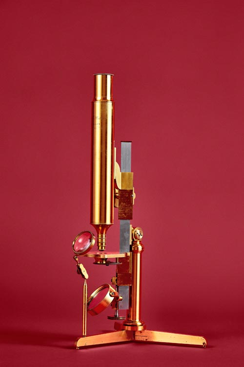
Carl Pistor and Friedrich Wilhelm Schiek, Berlin around 1830, the founders of cell theory, among others, appreciated the precision of the devices from this workshop. © Katharina Bohm, MDC
Progress in medicine and science would be unthinkable without microscopes. Today, structures can be visualized to the millionth of a millimeter, and living cell processes can be studied in fascinating detail. Based on historical microscopes from Berlin and Brandenburg, an exhibition at the MDC shows the path that has led to this point, demonstrating the enormous power of modern microscopes and their significance for medical research, both now and in the future.
On July 25, 2018, the permanent exhibition “Invisible – Visible – Transparent” officially opened at the MDC with a lecture event. The exhibition encompasses various objects, including some 30 historical microscopes from historic factories in Berlin and Brandenburg. These were assembled by Prof. Helmut Kettenmann, a neuroscientist at the MDC. He collected the historically significant microscopes and designed the exhibition, which bridges 19th century to present-day microscopy.
Where does microscopy stand today? At the opening ceremony, guest speaker Prof. Ernst Stelzer from Goethe University Frankfurt presented new optical microscope techniques that are used to produce three-dimensional images. But the development of microscopy and its applications are by no means at an end. Prof. Martin Lohse, Scientific Director of the MDC, anticipates promising developments in the field.
“At the MDC, a new microscopy center is currently being established with state-of-the-art technology. It will include new microscopy processes that go beyond the previous limits of microscopy in terms of sensitivity, speed, spatial resolution, and depth of cellular penetration.”
From amusing objects to lifesavers
The history of microscopy dates back to the 17th century, building on progress that had been made in the production of polished lenses for astronomy. Dutch lay scientist and fabric merchant Antoni van Leeuwenhoek (1632‒1723) constructed and used microscopes to visualize living structures such as blood cells. Up to the early 19th century, however, microscopes remained unique pieces which were mainly used for entertainment. Microscopy evenings were popular in the salons of high society; here, one could “regale the eyes” with “God’s smallest miracles.” Traveling microscopes were used for nature observation on excursions and outings.
“The triumph of the microscope in science began in the 19th century,” explains Prof. Helmut Kettenmann. The global center of microscopy was the burgeoning research metropolis Berlin. Some 80 microscope factories, the most well-known being Hartnack, Pistor, Schleiden, and Schieck, typically opened close to the university and its research institutes, cultivating active contacts with scientists in Berlin and around the world.
Famous Berlin scientists, including Johannes Müller, Rudolf Virchow, Robert Koch, and Hermann von Helmholtz, benefited from the flourishing microscopy industry while spurring it on with scientific discoveries. Microscopes from Berlin were used in countries like Italy and Spain, where they helped scientists such as Camillo Golgi (1843‒1926) and Ramón y Cajal (1852‒1943) discover brain cells and cellular structures. These achievements were honored with the Nobel Prize in 1906.
In the 19th century, the microscope became a lifesaving tool for meat inspections, which were initiated by the pathologist Rudolf Virchow in Berlin. Before further processing, pork was tested on-site for trichinella – parasites that are very dangerous to humans.
Microscopes find their way into lecture halls and schools
By 1900, microscopes were commonly used for scientific applications and increasingly permeated medical studies and school lessons. But the heyday of microscope manufacturing in Berlin and Brandenburg was already coming to an end. The lens system optimized by physicist Ernst Abbe shifted the focus of microscopy development to Jena, where the firm Carl Zeiss ousted the Berlin manufacturers from their leading position in the field.
The permanent exhibition “Invisible ‒ Visible ‒ Transparent” is on display on two floors of the MDC.C Conference Center. It is part of the Life Science Learning Lab Academy’s education program for secondary school students on the Berlin-Buch campus.
There are no regular opening times. If you are interested in visiting or participating in a tour of the exhibition, please contact the MDC Communications Department:
Email: communications@mdc-berlin.de
Phone: +49 (0)30 9406 2533
Dr. Annette Tuffs
Max-Delbrück-Centrum für Molekulare Medizin in der Helmholtz-Gemeinschaft
Leiterin Kommunikation
Phone: 030 – 9406 – 2140
annette.tuffs@mdc-berlin.de
Media Contact
More Information:
http://www.mdc-berlin.de/All latest news from the category: Event News
Newest articles

Parallel Paths: Understanding Malaria Resistance in Chimpanzees and Humans
The closest relatives of humans adapt genetically to habitats and infections Survival of the Fittest: Genetic Adaptations Uncovered in Chimpanzees Görlitz, 10.01.2025. Chimpanzees have genetic adaptations that help them survive…

You are What You Eat—Stanford Study Links Fiber to Anti-Cancer Gene Modulation
The Fiber Gap: A Growing Concern in American Diets Fiber is well known to be an important part of a healthy diet, yet less than 10% of Americans eat the minimum recommended…

Trust Your Gut—RNA-Protein Discovery for Better Immunity
HIRI researchers uncover control mechanisms of polysaccharide utilization in Bacteroides thetaiotaomicron. Researchers at the Helmholtz Institute for RNA-based Infection Research (HIRI) and the Julius-Maximilians-Universität (JMU) in Würzburg have identified a…


