Geology helps map kidney stone formation from tiny to troublesome

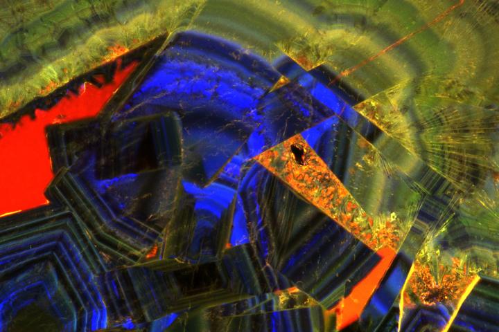
A fluorescent microscope image of a thin section of a human kidney stone reveals a complex history of crystal growth layering, fracturing, dissolution and recrystallization.
Image courtesy of Mayandi Sivaguru, University of Illinois
Advanced microscope technology and cutting-edge geological science are giving new perspectives to an old medical mystery: How do kidney stones form, why are some people more susceptible to them and can they be prevented?
In a new paper published in the journal Nature Reviews Urology, researchers from the University of Illinois Urbana-Champaign, Mayo Clinic and other collaborators described the geological nature of kidney stones, outlined the arc of their formation, established a new classification scheme and suggested possible clinical interventions.
“The process of kidney stone formation is part of the natural process of the stone formation seen throughout nature,” Illinois geology professor Bruce Fouke said. “We are bringing together geology, biology and medicine to map the entire process of kidney stone formation, step by step. With this road map in hand, more effective and targeted clinical interventions and therapies can now be developed.”
Kidney stones are a painful problem that will strike one in 10 adults in their lifetime and send half a million people in the United States to emergency rooms each year, according to the National Kidney Foundation. Yet little is understood about the geology behind how kidney stones form, Fouke said.
Previous work from Fouke’s group found that kidney stones form in the same way as geological stones in nature: Rather than crystalizing all at once, they partially dissolve and re-form multiple times, contrary to doctors’ belief that they form suddenly and intact.
In the new work, the research team – brought together by the Mayo Clinic and Illinois Alliance for Technology-Based Healthcare – describes in detail the multiple phases kidney stones go through in forming, dissolving and re-forming, documented through high-resolution imaging technologies. The findings defy the typical classification schemes doctors use, which are based on bulk analyses of the type of mineral and the presumed location of formation in the kidney. Instead, the researchers developed a new classification scheme based on which phase of formation the stone is in and which chemical processes it is undergoing.
“If we can identify these phase transformations, what makes one step to go to another and how it progresses, then perhaps we can intervene in that progression and break the chain of chemical reactions happening inside the kidney tissues before a stone becomes problematic,” said Mayandi Sivaguru, the lead author of the study and assistant director of core facilities at the Carl R. Woese Institute for Genomic Biology at Illinois.
One particularly revelatory finding was in the very beginnings of kidney stone formation: Stones start as microspherules, or tiny droplets of mineral, which merge to form larger crystals throughout kidney tissues. Normally they are flushed out, but when they coalesce together to form larger stones that continue to grow, they can become excruciatingly painful and even deadly in some cases, Fouke said.
“Stone formation is part of a natural, healthy process within kidneys where these tiny mineral deposits are shuttled away and excreted from the body,” Fouke said. “But then there is a tipping point when those same mineral deposits start to grow together too rapidly and are physically unable to leave the kidney.”
As the stone goes through the formation process, more microspherules merge, lose their rounded shape and transform into much larger, perfectly geometric crystals. Stones go through multiple cycles of partially dissolving – shedding up to 50% of their volume, the researchers found – and then growing again, creating a signature pattern of layered crystals much like those of agates, coral skeletons and hot-spring deposits seen around the world.
“Looking at a cross-section of a kidney stone, you would never guess that each of the layers was originally a bunch of little balls that lined up and coalesced. These are revolutionary new ways for us to understand how these minerals grow within the kidney and provide specific targets for stone growth prevention,” Fouke said.
The study authors outlined several possible clinical interventions and treatment targets based on this expanded knowledge of kidney stone formation. They hope that researchers and clinicians can explore and test these options, from drug targets to changes in diet or supplements that could disrupt the chemical and biological cascade driving kidney stone formation, Sivaguru said.
To aid in this testing, Fouke’s group developed the GeoBioCell, a microfluidic cartridge designed to mimic the intricate internal structures of the kidney. The group hopes the device can accelerate not only research, but also clinical diagnostic testing and the evaluation of potential therapies, especially for the more than 70% of kidney stone patients with recurring stones.
“Ultimately, our vision is that every operating room would have a small geology lab attached. In that lab, you could do a very rapid diagnostic on a stone or stone fragment in a matter of minutes, and have informed and individualized treatment targets,” Fouke said.
###
The Mayo-Illinois Alliance, the Mayo Clinic Center for Individualized Medicine, the Mayo Clinic O’Brien Urology Research Center and the Astrobiology Institute of the National Aeronautics and Space Administration supported this work.
Editor’s note: To reach Bruce Fouke, call 217-244-5431; email fouke@illinois.edu. To reach Mayandi Sivaguru, email sivaguru@illinois.edu.
The paper “Human kidney stones: A natural record of universal biomineralization” is available online and from the U. of I. News Bureau.Editor’s note: To reach Bruce Fouke, call 217-244-5431; email fouke@illinois.edu. To reach Mayandi Sivaguru, email sivaguru@illinois.edu.
The paper “Human kidney stones: A natural record of universal biomineralization” is available online and from the U. of I. News Bureau.
Media Contact
All latest news from the category: Health and Medicine
This subject area encompasses research and studies in the field of human medicine.
Among the wide-ranging list of topics covered here are anesthesiology, anatomy, surgery, human genetics, hygiene and environmental medicine, internal medicine, neurology, pharmacology, physiology, urology and dental medicine.
Newest articles

Innovative 3D printed scaffolds offer new hope for bone healing
Researchers at the Institute for Bioengineering of Catalonia have developed novel 3D printed PLA-CaP scaffolds that promote blood vessel formation, ensuring better healing and regeneration of bone tissue. Bone is…

The surprising role of gut infection in Alzheimer’s disease
ASU- and Banner Alzheimer’s Institute-led study implicates link between a common virus and the disease, which travels from the gut to the brain and may be a target for antiviral…

Molecular gardening: New enzymes discovered for protein modification pruning
How deubiquitinases USP53 and USP54 cleave long polyubiquitin chains and how the former is linked to liver disease in children. Deubiquitinases (DUBs) are enzymes used by cells to trim protein…


