Inselspital: Images from the depths of the brain

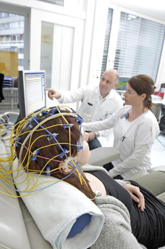
The new method can do what the surface EEG cannot: show electrical activities in the brain. Inselspital, Bern University Hospital
For over 20 years, doctors have been dreaming of depicting the brain’s electrical activity in an MRI scan. Researchers from Bern University Hospital have now succeeded in making this dream a reality by means of a unique method.
The electrical fields are measured indirectly through their effect on magnetic fields,rather than directly. But although a healthy brain’s electrical fields are too weak to produce a measurable disruption of the magnetic field, neuroradiologists can now measure this where the fields are more pronounced in the short term. In patients with epilepsy.
Imaging helps to localise epilepsy and shows healing
The research team of physicist Dr Claus Kiefer and doctors Eugenio Abela, Kaspar Schindler and Roland Wiest from the Support Center for Advanced Neuroimaging (SCAN) at the Department of Diagnostic and Interventional Neuroradiology and the Department of Neurology at Bern University Hospital used the new method in a pilot study with eight epilepsy patients.
In doing so, it was found that the newly developed MR sequence makes magnetic field disturbances visible, even in deep regions of the brain. The surface EEG, which is otherwise used, has never achieved this. As a result, it is possible to localise the origin of the epileptic seizures with even more precision, which benefits those patients who exhibit no structural abnormalities in the “normal” MRI.
In addition, researchers demonstrated that epilepsy patients, who are seizure-free after an operation, no longer show such magnetic field disturbances – that their brain works “disruption-free” like that of a healthy person. On the other hand, patients who continued to have seizures still demonstrated the typical pathological signals. These astounding new insights into the function of our brain were published on 29th January in the renowned American journal, Radiology.
Patented methods only in Bern
The revolutionary imaging method is patented by the University of Bern and is only currently offered at Bern University Hospital. The advantage: If epilepsy patients, whose medication is not helping, need an MRI examination, just eight additional minutes in the MRI can better localise the region of origin of the exaggerated electrical brain activity. This is also the case if patients are not currently having a seizure, as the method is so sensitive that it even records weak epileptic activity that exists between the actual seizures.
The newly developed MR sequence is now expected to be validated internationally in further clinical studies.
Contact:
Prof Dr Roland Wiest, Chief Physician, Department of Diagnostic and Interventional Neuroradiology, Bern University Hospital, +41 31 632 36 73.
Prof Dr Kaspar Schindler, Chief Physician, Department of Neurology, Bern University Hospital, +41 31 632 30 54.
http://www.ncbi.nlm.nih.gov/pubmed/26824710
http://www.neurorad.insel.ch/
Media Contact
All latest news from the category: Health and Medicine
This subject area encompasses research and studies in the field of human medicine.
Among the wide-ranging list of topics covered here are anesthesiology, anatomy, surgery, human genetics, hygiene and environmental medicine, internal medicine, neurology, pharmacology, physiology, urology and dental medicine.
Newest articles

First-of-its-kind study uses remote sensing to monitor plastic debris in rivers and lakes
Remote sensing creates a cost-effective solution to monitoring plastic pollution. A first-of-its-kind study from researchers at the University of Minnesota Twin Cities shows how remote sensing can help monitor and…

Laser-based artificial neuron mimics nerve cell functions at lightning speed
With a processing speed a billion times faster than nature, chip-based laser neuron could help advance AI tasks such as pattern recognition and sequence prediction. Researchers have developed a laser-based…

Optimising the processing of plastic waste
Just one look in the yellow bin reveals a colourful jumble of different types of plastic. However, the purer and more uniform plastic waste is, the easier it is to…


