Novel bone imaging approach provides insights into the progression of knee osteoarthritis

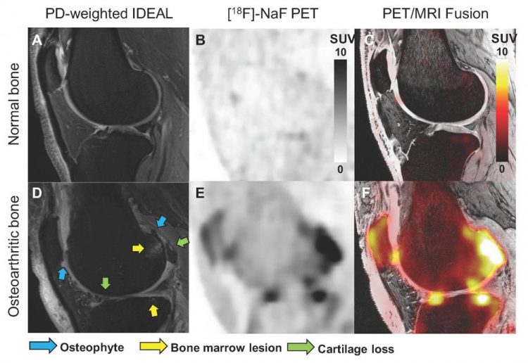
Representative structural MRI image (PD-weighted IDEAL image, A and D), [18F]NaF PET SUV images (B and E), and PET/MR fusion images (C and F) of a healthy knee and a knee with osteoarthritis. MOAKS scoring of MR images was used to identify the size of osteophytes, bone marrow lesions and cartilage loss within various bone regions in the patella, tibia and femur. Arrows highlight bone regions with osteophytes in blue, bone marrow lesions in yellow, and cartilage loss in green. Credit: LE Watkins, et al., Stanford University, CA.
Osteoarthritis, the most common joint disorder in the United States, affects more than 32.5 million adults. It is marked by degradation and loss of soft tissues, such as cartilage, and development of bone marrow lesions and osteophytes.
Osteoarthritis occurs most frequently in the hands, hips and knees and results in reduced productivity and quality of life.
“Osteoarthritis is not well understood, in part because we lack the tools to objectively evaluate early and reversible changes in key tissues,” noted Lauren Watkins, MS, a researcher at the Stanford University Imaging of Musculoskeletal Function Group in Stanford, California.
“While many MRI methods have been developed for assessment of early degenerative changes in cartilage, functional imaging of bone in the joint remains a major challenge.”
To evaluate the relationship between structural and physiological changes in knee osteoarthritis, researchers utilized positron emission tomography (PET)/MRI with 18F NaF to image both knees of 12 subjects. Five subjects were scanned twice, with at least five days between visits, to assess repeatability of the technique.
MRI Osteoarthritis Knee Score (MOAKS) assessment of each knee was performed by a trained musculoskeletal radiologist, and dynamic PET data were used to calculate the rates of bone perfusion, tissue clearance and mineralization, as well as tracer extraction fraction and total bone uptake rate.
Kinetic modeling was performed for regions of interest representing the subchondral bone of the patella, medial and lateral tibia, and anterior, central, and posterior regions of the medial and lateral femur.
The knees with MOAKS findings were divided into regions of cartilage loss, bone marrow lesions and osteophytes and were analyzed along with the kinetic parameters derived from PET data.
Abnormal bone metabolism in regions with bone marrow lesions, osteophytes, and adjacent cartilage lesions was found to be strongly associated with greater bone perfusion rates as compared to bone that appeared normal on MRI. Additionally, strong spatial relationships between bone metabolic abnormalities and changes in overlying cartilage were noted.
“These findings show the utility and potential of PET imaging to study the role of bone physiology in degenerative joint disease,” said Watkins. “This knowledge may help us understand the order of events leading to structural and functional degeneration of the knee. Further, this will help us to develop and quickly evaluate new interventions that target specific metabolic pathways to give us the best chance to slow or arrest the onset and progression of osteoarthritis.”
###
Abstract 182. “Evaluating the Relationship between Dynamic Na[18F]F-Uptake Parameters and MRI Knee Osteoarthritic Findings,” Lauren Watkins, Valentina Mazzoli, Scott Uhlrich, Garry Gold and Feliks Kogan, Stanford University, Stanford, California; James MacKay, University of Cambridge, Cambridge, United Kingdom; and Bryan Haddock, Rigshospitalet, Copenhagen, Denmark. SNMMI's 67th Annual Meeting, July 11-14, 2020.
All 2020 SNMMI Annual Meeting abstracts can be found online at http://jnm.
About the Society of Nuclear Medicine and Molecular Imaging
The Society of Nuclear Medicine and Molecular Imaging (SNMMI) is an international scientific and medical organization dedicated to advancing nuclear medicine and molecular imaging, vital elements of precision medicine that allow diagnosis and treatment to be tailored to individual patients in order to achieve the best possible outcomes.
SNMMI's members set the standard for molecular imaging and nuclear medicine practice by creating guidelines, sharing information through journals and meetings and leading advocacy on key issues that affect molecular imaging and therapy research and practice. For more information, visit http://www.
Media Contact
All latest news from the category: Health and Medicine
This subject area encompasses research and studies in the field of human medicine.
Among the wide-ranging list of topics covered here are anesthesiology, anatomy, surgery, human genetics, hygiene and environmental medicine, internal medicine, neurology, pharmacology, physiology, urology and dental medicine.
Newest articles

First-of-its-kind study uses remote sensing to monitor plastic debris in rivers and lakes
Remote sensing creates a cost-effective solution to monitoring plastic pollution. A first-of-its-kind study from researchers at the University of Minnesota Twin Cities shows how remote sensing can help monitor and…

Laser-based artificial neuron mimics nerve cell functions at lightning speed
With a processing speed a billion times faster than nature, chip-based laser neuron could help advance AI tasks such as pattern recognition and sequence prediction. Researchers have developed a laser-based…

Optimising the processing of plastic waste
Just one look in the yellow bin reveals a colourful jumble of different types of plastic. However, the purer and more uniform plastic waste is, the easier it is to…


