Seeing the Knee in a New Light: Fluorescent Probe Tracks Osteoarthritis Development

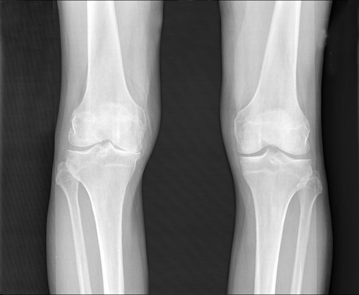
National Institute of Arthritis and Musculoskeletal and Skin Diseases (NIAMS) X-ray image of osteoarthritic knees.
A fluorescent probe may make it easier to diagnose and monitor osteoarthritis, a painful joint disease affecting nearly 27 million Americans. The disease is often detected late in development after painful symptoms occur. Earlier diagnosis might lead to better management and patient outcomes. A new study reports that a fluorescent probe tracked the development of osteoarthritis in male mice, brightening as the disease progressed. The findings are published in the February issue of Arthritis & Rheumatology.
In this case, the “probe” was a harmless fluorescent molecule that detected the activity leading to cartilage loss in the joint, the key characteristic of osteoarthritis. This lab and mouse study, led by researchers from Tufts University School of Medicine (TUSM) and the Sackler School of Graduate Biomedical Sciences at Tufts, is the first to demonstrate that near-infrared fluorescence – a specific type of light invisible to the human eye but possible to see with optical imaging – can be used to detect osteoarthritis changes over time. The new approach might also help analyze the effectiveness of osteoarthritis drugs, leading to improved treatments.
“Patients are frequently in pain by the time osteoarthritis is diagnosed. The imaging tests most frequently used, X-rays, don’t indicate the level of pain or allow us to directly see the amount of cartilage loss, which is a challenge for physicians and patients,” said co-first author Averi A. Leahy, B.A., an M.D./Ph.D. student in the medical scientist training program at TUSM and the Sackler School.
“The fluorescent probe made it easy to see the activities that lead to cartilage breakdown in the initial and moderate stages of osteoarthritis, which is needed for early detection and adequate monitoring of the disease. To measure the probe’s signal, we used an optical imaging system, to non-invasively look inside the knee,” said co-first author Shadi A. Esfahani, M.D., M.P.H., post-doctoral fellow in the division of nuclear medicine and molecular imaging at Massachusetts General Hospital, and in the department of radiology at Harvard Medical School.
The right knees of 54 mice were affected by injury-induced osteoarthritis and served as the experimental group (the mice received pain medication). The healthy, left knees of the mice served as the control group.
Over a two-month period, the researchers took images of each knee every two weeks to determine if the fluorescent probe emitted a signal. Strikingly, the signal became brighter in the injured right knee, at every examined time point, through the early to moderate stages of osteoarthritis. The probe emitted a lower signal in the healthy left knee, and did not increase significantly over time.
The corresponding and senior author is Li Zeng, Ph.D., associate professor in the department of integrative physiology and pathobiology at TUSM, and member of the cellular, molecular and developmental biology program faculty at the Sackler School. Averi Leahy is one of her students. The work was done in collaboration with Umar Mahmood, M.D., Ph.D., director of the Center for Translational Nuclear Medicine and Molecular Imaging, co-director of Nuclear Medicine and Molecular Imaging, both at Massachusetts General Hospital; and associate professor in the department of radiology at Harvard Medical School.
Zeng reports that the next step is to monitor the fluorescent probe over a longer period of time to determine whether the same results are produced during the late stages of osteoarthritis. She also hopes to use the probe to help develop treatments for animals, as some dogs are prone to osteoarthritis.
Osteoarthritis, the most common type of arthritis, causes pain, swelling, and reduced motion by breaking down cartilage that covers the ends of bones. The disease often results from an injury or long term wear and tear to the hip, hands, or knees. Reducing the burden of osteoarthritis has been a top priority for US public health officials. The National Public Health Agenda for Osteoarthritis, created by the Arthritis Foundation and the Centers for Disease Control and Prevention, calls for more research into osteoarthritis to better understand effective strategies for intervention.
The Zeng laboratory at Tufts studies the mechanisms of tissue formation and degradation to develop effective strategies to treat arthritis. Her team uses tissue engineering techniques to examine approaches that can be used to generate stronger, more stable cartilage tissue that is resistant to inflammation commonly caused by osteoarthritis.
Previous research had identified that an increase in enzymes called matrix metalloproteinases (MMP) contributes to the cartilage degradation that occurs with osteoarthritis. The fluorescent probe was introduced into the knee joint and measured MMP activity.
Additional authors of this study are Carrie K. Hui, B.S., a doctoral student in the cellular, molecular and developmental biology program at the Sackler School; Roshni S. Rainbow, Ph.D., an affiliate in the department of anatomy and basic sciences at Tufts University School of Medicine and former Institutional Research and Academic Career Development (IRACDA) postdoctoral fellow at the Sackler School; Daisy S. Nakamura, B.S., a doctoral student in the cellular, molecular and developmental biology program at the Sackler School; and Brian H. Tracey, Ph.D., professor in the department of electrical and computer engineering at Tufts University School of Engineering. Research reported in this publication was supported by the National Institute of Arthritis and Musculoskeletal and Skin Diseases of the National Institutes of Health (NIH) under award numbers F30AR065866 and R01AR059106, by a research training grant from the National Institute of General Medical Sciences (NIGMS) of the NIH under award number K12GM074869 (IRACDA), and by the National Center for Advancing Translational Sciences of the NIH under award number UL1TR001064. Additional funding was provided by a Tufts Collaborates seed grant, and the National Science Foundation under award number CBET-0966920.
The Institutional Research Career and Academic Development Awards from the NIGMS combine traditional post-doctoral research work with mentored teaching experience at institutions that serve under-represented minorities. The Sackler School’s IRACDA Training in Education and Critical Research Skills (TEACRS) program is a partnership with the University of Massachusetts, Boston; Pine Manor College, and Bunker Hill Community College. It is the only IRACDA-funded program in New England.
Leahy, A. A., Esfahani, S. A., Foote, A. T., Hui, C. K., Rainbow, R. S., Nakamura, D. S., Tracey, B. H., Mahmood, U., & Zeng, L. (February 2015). “Analysis of the Trajectory of Osteoarthritis Development in a Mouse Model by Serial Near-Infrared Fluorescence Imaging of Matrix Metalloproteinase Activities.” Arthritis & Rheumatology, 67(2). DOI: 10.1002/art.38957
About Tufts University School of Medicine and the Sackler School of Graduate Biomedical Sciences
Tufts University School of Medicine and the Sackler School of Graduate Biomedical Sciences at Tufts University are international leaders in innovative medical and population health education and advanced research. Tufts University School of Medicine emphasizes rigorous fundamentals in a dynamic learning environment to educate physicians, scientists, and public health professionals to become leaders in their fields. The School of Medicine and the Sackler School are renowned for excellence in education in general medicine, the biomedical sciences, and public health, as well as for innovative research at the cellular, molecular, and population health level. Ranked among the top in the nation, the School of Medicine is affiliated with six major teaching hospitals and more than 30 health care facilities. Tufts University School of Medicine and the Sackler School undertake research that is consistently rated among the highest in the nation for its effect on the advancement of medical and prevention science.
Contact Information
Siobhan Gallagher
Associate Director, Public Relations
siobhan.gallagher@tufts.edu
Phone: 617-636-6586
Media Contact
More Information:
http://www.tufts.edu/All latest news from the category: Health and Medicine
This subject area encompasses research and studies in the field of human medicine.
Among the wide-ranging list of topics covered here are anesthesiology, anatomy, surgery, human genetics, hygiene and environmental medicine, internal medicine, neurology, pharmacology, physiology, urology and dental medicine.
Newest articles

Innovative vortex beam technology
…unleashes ultra-secure, high-capacity data transmission. Scientists have developed a breakthrough optical technology that could dramatically enhance the capacity and security of data transmission (Fig. 1). By utilizing a new type…

Tiny dancers: Scientists synchronise bacterial motion
Researchers at TU Delft have discovered that E. coli bacteria can synchronise their movements, creating order in seemingly random biological systems. By trapping individual bacteria in micro-engineered circular cavities and…

Primary investigation on ram-rotor detonation engine
Detonation is a supersonic combustion wave, characterized by a shock wave driven by the energy release from closely coupled chemical reactions. It is a typical form of pressure gain combustion,…


