3-dimensionally images in magnetic resonance

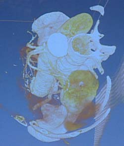
In the near future, images obtained from magnetic resonance will be common. The aim of the TRAC project is to be able to see internal organs 3-dimensionally using a non-invasive technique. Currently images of the liver are being worked with, but it is hoped that the technique will be useful for any internal structure or tissue.
Vicomtech is one of the enterprises located in the Miramón technological Park. Here they work with, amongst other things, computer-treated images. One of the lines of research currently being undertaken is medical applications and it is here the TRAC project becomes involved. For the moment they are working on the treatment of images of the liver.
The aim of the TRAC project is the creation of a prototype based on reality techniques which enable doctors to obtain 3-D images of patients’ internal organs. The idea is that the prototype will help in planning and developing operations.
In magnetic resonance, an image of the liver is obtained in cross-section, normally every three millimetres. These bidimensional images are printed separately on to acetate and it is the medical consultant, on examining them, who makes a mental representation of the volume of the liver, in three-dimensional terms, based on her or his knowledge and experience.
In order to create images directly in three dimensions, one starts with the two-dimensional ones obtained with the resonance. First, by means of a computer, filters are applied in order to eliminate noise and to highlight the principal features; the right filter has to be applied, because it is also necessary to see to the most important details. Then mathematical segmentation algorithms are applied. The aim of the segmentation is to identify and lift the liver surface from the resonance images. This is the key step in the process, given that the liver is a large and complex organ. Finally, reconstruction algorithms in three dimensions are applied to these results.
With these images the doctors can study the external anatomy of the patient’s liver in more detail and the extent of the damage. Moreover, when the capacity for detail of the system is increased, the liver interior may be explored; blood vessels, tumours, etc.
The project has a second part which employs reality techniques carried out in the operating theatre. The aim of this is to see the operating theatre. For this to happen, a number of steps have to be followed.
The development and research for all this project is being undertaken by Vicomtech, but there are also collaborating bodies: STT, which develops and markets monitoring systems; BILBOMATICA, specialists in the handling of medical data bases, and finally, the results are supervised by a group of specialist from the Hospital de Cruces (Bilbao) for their verification and usefulness. It is a team of doctors which give the green light, as it were, for the product.
The best thing, without any doubt, is not to have the need to carry out a scanner operation and doctors will be able to have much more detailed information once the project has been developed.
Media Contact
All latest news from the category: Information Technology
Here you can find a summary of innovations in the fields of information and data processing and up-to-date developments on IT equipment and hardware.
This area covers topics such as IT services, IT architectures, IT management and telecommunications.
Newest articles

Global Genetic Insights into Depression Across Ethnicities
New genetic risk factors for depression have been identified across all major global populations for the first time, allowing scientists to predict risk of depression regardless of ethnicity. The world’s…

Back to Basics: Healthy Lifestyle Reduces Chronic Back Pain
Low back pain is a leading cause of disability worldwide with many treatments, such as medication, often failing to provide lasting relief. Researchers from the University of Sydney’s Centre for Rural…

Retinoblastoma: Eye-Catching Investigation into Retinal Tumor Cells
A research team from the Medical Faculty of the University of Duisburg-Essen and the University Hospital Essen has developed a new cell culture model that can be used to better…


