Different forms of cellular adhesion structures can interconvert

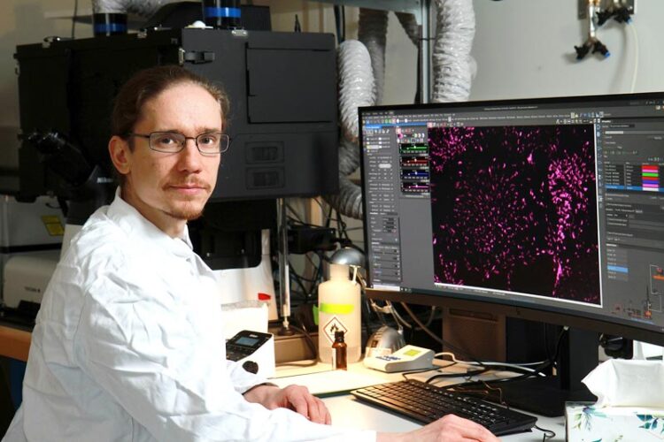
Dr Fabian Lukas researches adhesion structures in cells. Here you can see a microscope image of human pigment epithelial cells in which the integrin is fluorescently labelled.
Copyright: RPTU, Koziel
Cells form adhesion structures to anchor themselves in their environment. The coordinated assembly and disassembly of these adhesions also enables cells to move from one place to another. There are various forms of adhesions. Focal adhesions are the best-studied type. Until now, they were believed to be always built up anew when cells move. A study led by a team of researchers from Kaiserslautern has now shown for the first time that different forms of adhesions can interconvert. During this process a protein scaffold remains intact. Only the proteins bound to it change, as the team writes in the current issue of the journal Nature Communications.
There are cells in our body that are densely anchored within tissues and other cells that move like immune cells. They all have in common that they require certain structures to adhere to their environment. “These are special protein complexes that make adhesion possible,” says Professor Dr. Tanja Maritzen, who conducts research into nanophysiology at the University of Kaiserslautern-Landau (RPTU).
Such adhesions not only play a role in cells within a tissue, but also in processes in which cells have to move, for example during embryonic development or when cells have to migrate to close wounds. They are also important for the communication of cells with their environment. “This means that under certain conditions cells need very long-lasting adhesions, while under other conditions dynamic structures are required to enable locomotion,” continues the professor. Accordingly, researchers distinguish between different types of adhesion structures: “Focal adhesions, also known as canonical adhesion, are the ones that have been best-studied.”
A specific protein complex in the cell membrane is responsible for this form of adhesion. It is structured as follows: Special proteins, the integrins, are anchored in the membrane. They have a part outside of the cell with which they bind to specific proteins of the extracellular matrix and thus adhere to the material cells are embedded in. The integrins are also firmly attached to structures inside the cell via a protein complex. This contains, for example, paxillin as a typical component of focal adhesions.
In addition, there are so-called reticular adhesions, adhesion networks and retraction fibres, all of which also contain integrins, but otherwise differ in their composition, for example, no paxillin is found in them. These three types of adhesions are also referred to as non-canonical adhesions. They have not yet been well studied.
“Until now, it was believed that focal adhesions arise completely anew, e.g. when cells move,” continues Maritzen. In their current study, the team led by the Kaiserslautern professor and her colleague Dr. Fabian Lukas investigated the question of whether the different forms of adhesions can instead also convert into each other. Lukas, the first author of the current study, explains. “We hypothesised that the integrins remain intact as the basic scaffold while the associated molecular complexes are exchanged.”
In their investigations, the research group has benefited from the fact that they have been working with a specific protein, Stonin1, for a long time. “This protein is found in non-canonical adhesions, but not in focal adhesions,” explains Lukas, “and can therefore be used as an identification feature for these structures.”
To test their hypothesis, the researchers carried out a series of experiments. For this, they modified the genes for stonin1 and integrin ß5 with the CRISPR/Cas9 gene scissors, attaching the DNA sequence for a fluorescent protein to one end. This makes it possible to observe them in the cell using fluorescence microscopy. In addition, they labelled paxillin.
They then looked at the adhesion structures on membranes of living cells using a high-resolution microscope and followed their development, e.g. while a cell is dividing. For division, the cell has to form into a sphere, disassembling its focal adhesions in the process. “Such a cell cycle takes around 120 minutes. During this time, we have seen that the integrins remain unchanged,” explains Lukas.
However, the situation was different for the proteins paxillin and stonin1. “We observed that paxillin disappears over time, while stonin1 appears. The adhesion structures are therefore still present in the cell, they just change their molecular composition,” concludes Maritzen.
During cell division, the cell uses reticular adhesions to attach to its environment. After division, the following can be observed: In the two daughter cells, the reticular adhesions become focal adhesions again.
In a further experiment, they investigated what happens in cells that are in motion. “The cells leave behind membrane strands, so-called retraction fibres, when they migrate. Here, too, we saw that integrins remain in these structures as a stable scaffold. When the cell changes direction and moves back across the retraction fibres, stonin1 is replaced by paxillin over time, so that a retraction fibre becomes a focal adhesion,” summarises Lukas.
The results show for the first time a close connection between the different forms of adhesions: Focal adhesions do not always arise from scratch, as previously assumed, but also through recycling of a stable integrin backbone in which only specific binding partners are exchanged.
Researchers from the RPTU in Kaiserslautern, the Leibniz-Forschungsinstitut für Molekulare Pharmakologie in Berlin, the Max Delbrück Centre for Molecular Medicine in Berlin, the Freie Universität Berlin, Charité Universitätsmedizin and the National Heart, Lung, and Blood Institute in Bethesda (Maryland) were involved in the study.
The study has been published in the renowned journal Nature Communications: “Canonical and non-canonical integrin-based adhesions dynamically interconvert”.
DOI: https://doi.org/10.1038/s41467-024-46381-x
Questions can be adressed to:
Professorin Dr. Tanja Maritzen
Nanophysiology
RPTU in Kaiserslautern
E-Mail: maritzen[at]rptu.de
Tel.: +49 (0)631 205-4908
Wissenschaftliche Ansprechpartner:
Professorin Dr. Tanja Maritzen
Nanophysiology
RPTU in Kaiserslautern
E-Mail: maritzen[at]rptu.de
Tel.: +49 (0)631 205-4908
Originalpublikation:
Nature Communications
“Canonical and non-canonical integrin-based adhesions dynamically interconvert”
DOI: https://doi.org/10.1038/s41467-024-46381-x
Media Contact
All latest news from the category: Life Sciences and Chemistry
Articles and reports from the Life Sciences and chemistry area deal with applied and basic research into modern biology, chemistry and human medicine.
Valuable information can be found on a range of life sciences fields including bacteriology, biochemistry, bionics, bioinformatics, biophysics, biotechnology, genetics, geobotany, human biology, marine biology, microbiology, molecular biology, cellular biology, zoology, bioinorganic chemistry, microchemistry and environmental chemistry.
Newest articles

Innovative 3D printed scaffolds offer new hope for bone healing
Researchers at the Institute for Bioengineering of Catalonia have developed novel 3D printed PLA-CaP scaffolds that promote blood vessel formation, ensuring better healing and regeneration of bone tissue. Bone is…

The surprising role of gut infection in Alzheimer’s disease
ASU- and Banner Alzheimer’s Institute-led study implicates link between a common virus and the disease, which travels from the gut to the brain and may be a target for antiviral…

Molecular gardening: New enzymes discovered for protein modification pruning
How deubiquitinases USP53 and USP54 cleave long polyubiquitin chains and how the former is linked to liver disease in children. Deubiquitinases (DUBs) are enzymes used by cells to trim protein…


