More effective cell studies using new AI method

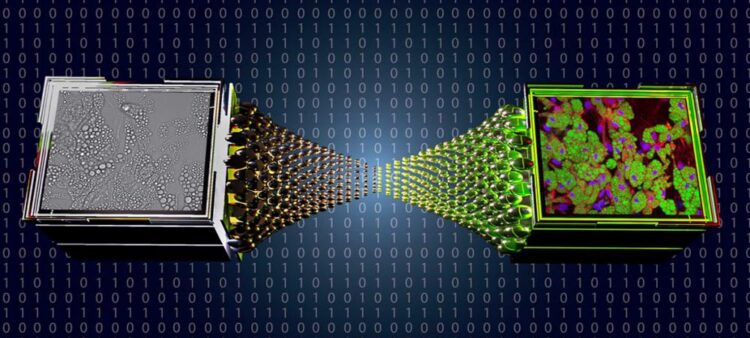
Virtual staining image
https://www.expertsvar.se/wp-content/uploads/2021/10/virtual_staining_image_1200-1024x576.jpg
A new study from the University of Gothenburg opens the way for more effective microscopy, making it easier to research diseases. The study shows how artificial intelligence can be used to develop faster, cheaper and more reliable information about cells, while also eliminating the disadvantages from using chemicals in the process.
Studying cells and their components is a cornerstone of biomedicine and pharmaceutical research and can provide information about the health of cells, responses to different medications or deviations in the cell structure.
Two of the most common methods for studying cells using microscopes (bright field microscopy and fluorescence microscopy) both have advantages and disadvantages. Bright field microscopy, where the cell is illuminated with a bright light, is a simple and quick method, but it cannot accentuate individual cell components to provide specific targeted information about the cell. This, however, is possible with fluorescence microscopy, where part of the cell is stained with a substance that stands out under the microscope.
Images equivalent to fluorescence images – without chemicals
At the same time, fluorescence microscopy has many disadvantages, which a research team at the University of Gothenburg has now addressed.
“Fluorescence microscopy is effective for studying cells, since the method is very precise in accentuating the most interesting information. The problem is that it is expensive, time consuming and complicated to stain cells, while the chemicals in the stain risk damaging the cells or inhibiting the processes being studied. This is why we developed a method to create the same process digitally, so that we get all the advantages of fluorescence microscopy without its disadvantages,” says Jesús Pineda, a doctoral student in physics at the University of Gothenburg.
Together with Saga Helgadottir and Benjamin Midtvedt, Pineda is the main author of the recently published study examining how deep learning – a form of artificial intelligence (AI) – can be used to translate bright field images to equivalent fluorescence images.
Simpler, more reliable and cheaper method
With this new AI-based method, it is possible to use an image made with a bright field microscope and calculate how the same image would look if it had been taken with a fluorescence microscope.
“This means it is easier, cheaper and less time-consuming to extract important information about cells. The results are more reliable since chemicals do not need to be added, and the cells can be followed over time since they are not damaged. The method provides more reproducible results so that results from different labs can more easily be compared.”
Can facilitate hospital analyses
For the moment, the researchers want to continue developing the method, but, ultimately, they see huge potential for hospitals to utilise the findings.
“It would really be helpful if hospitals could avoid chemical staining during microscopy and produce quicker and more reliable test results at a lower cost. The method is also particularly suitable for hospital laboratories, where you often want to test the same type of samples repeatedly.”
Wissenschaftliche Ansprechpartner:
Contact:
Jesús Pineda, doctoral student in physics at the University of Gothenburg, phone: 0046 737 360458, e-mail jesus.pineda@physics.gu.se
Benjamin Midtvedt, doctoral student in physics at the University of Gothenburg, phone: 0046 730 752304, e-mail: benjamin.midtvedt@physics.gu.se
Saga Helgadottir, post-doctoral researcher in physics at the University of Gothenburg, phone: 0046 722 769079, e-mail: saga.helgadottir@physics.gu.se
Originalpublikation:
Scientific magazine: Biophysics reviews
Title: Extracting quantitative biological information from bright-field cell images using deep learning
Weitere Informationen:
https://aip.scitation.org/doi/10.1063/5.0044782
https://www.expertsvar.se/wp-content/uploads/2021/10/virtual_staining_image_1200…
Media Contact
All latest news from the category: Life Sciences and Chemistry
Articles and reports from the Life Sciences and chemistry area deal with applied and basic research into modern biology, chemistry and human medicine.
Valuable information can be found on a range of life sciences fields including bacteriology, biochemistry, bionics, bioinformatics, biophysics, biotechnology, genetics, geobotany, human biology, marine biology, microbiology, molecular biology, cellular biology, zoology, bioinorganic chemistry, microchemistry and environmental chemistry.
Newest articles

Parallel Paths: Understanding Malaria Resistance in Chimpanzees and Humans
The closest relatives of humans adapt genetically to habitats and infections Survival of the Fittest: Genetic Adaptations Uncovered in Chimpanzees Görlitz, 10.01.2025. Chimpanzees have genetic adaptations that help them survive…

You are What You Eat—Stanford Study Links Fiber to Anti-Cancer Gene Modulation
The Fiber Gap: A Growing Concern in American Diets Fiber is well known to be an important part of a healthy diet, yet less than 10% of Americans eat the minimum recommended…

Trust Your Gut—RNA-Protein Discovery for Better Immunity
HIRI researchers uncover control mechanisms of polysaccharide utilization in Bacteroides thetaiotaomicron. Researchers at the Helmholtz Institute for RNA-based Infection Research (HIRI) and the Julius-Maximilians-Universität (JMU) in Würzburg have identified a…


