New methods reveal the biomechanics of blood clotting

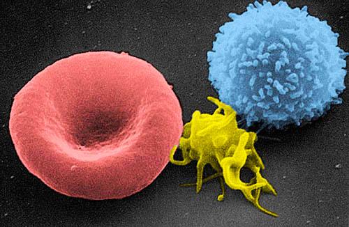
A scanning electron micrograph shows a red blood cell, an activated platelet (in yellow) and a white blood cell. The ability to map the magnitude and orientation of forces on a cell provides a new tool for investigating not just blood clotting but a range of biomechanical processes. Credit: National Cancer Institute
Platelets are cells in the blood whose job is to stop bleeding by sticking together to form clots and plug up a wound. Now, for the first time, scientists have measured and mapped the key molecular forces on platelets that trigger this process.
The extensive results are published in two separate studies, in the Proceedings of the National Academy of Sciences (PNAS) and in Nature Methods.
“We show conclusively that, in order to activate clotting, the cell needs a targeted force of a magnitude of just a few piconewtons — or a force about a billion times less than the weight of a staple,” says Khalid Salaita, associate professor in Emory University's Department of Chemistry and the lead author of the studies. “The real surprise we found is that platelets care about the direction of that force and that it has to be lateral. They're very picky. But they should be picky because otherwise they might accidentally create a clot. That's what causes strokes.”
Fibrinogen, the third most abundant protein in blood, acts like glue to stick platelets together as a clot forms. Each platelet has about 70,000 copies of a receptor for fibrinogen on its surface. These receptors can work like grappling hooks to latch onto fibrinogen.
“What was puzzling,” Salaita explains, “is that platelets, despite having all these receptors, do not normally latch onto the abundant fibrinogen. They keep flowing past it until you have an injury and fibrinogen becomes anchored. Then the platelets rapidly bind to fibrinogen allowing platelets to aggregate and for clotting to proceed.”
The Salaita lab is a leader in visualizing and mapping the mechanical forces applied by cells. In order to explore the biomechanics of blood clotting, the lab teamed up with physician and biomedical engineer Wilbur Lam, an expert in hematology at Emory's School of Medicine. Both Salaita and Lam are also affiliated with Emory's Winship Cancer Institute and the Wallace H. Coulter Department of Biomedical Engineering at Emory and Georgia Tech.
In initial experiments, for the PNAS paper, the Salaita lab anchored fibrinogen ligands onto a lipid membrane. On this surface, the ligands could slip and slide laterally, but resisted motion perpendicular to the surface — similar to the way a hockey puck slides easily over the surface of an ice rink but is harder to lift off of the plane of ice. The researchers then introduced platelets to this surface and experiments showed that the platelets failed to activate and stick together.
In contrast, when the fibrinogen ligands were anchored to a glass slide and unable to move laterally, the platelets rapidly activated. Using tension-imaging technology it developed, the Salaita lab showed that the platelets applied forces between five and 20 piconewtons to initiate activation.
“Platelets have to walk this tightrope between stopping bleeding quickly and accurately during an injury but avoiding unnecessary clotting. Mistakes could be fatal,” Salaita says. “We think they use this lateral force signal like a safety lock to prevent unnecessary clotting.”
Blood vessels are lined with endothelial cells and an injury exposes the fibrous matrix underneath these cells, Salaita explains. Platelets and fibrinogen in the blood can then stick to the injury site.
Salaita theorizes that when a platelet encounters stuck fibrinogen molecules, the platelet tugs on this fibrinogen as a way to test it. The resulting force generates a potent signal to activate platelets and that allows them to grab the fibrinogen from the blood, driving the process of clumping with other platelets.
The abnormal clotting that leads to strokes, and the uncontrollable bleeding of hemophilia, may be related to malfunctions in this biomechanical mechanism, he adds.
In 2011, the Salaita lab developed a fluorescence-sensor method for mapping cell mechanics. Alexa Mattheyses, a cell biologist at Emory's School of Medicine and Winship Cancer Institute, teamed with the lab to test whether fluorescence polarization could be applied to map the direction of cell forces and provide further insights into the biomechanics of blood clotting.
The results, published in the Nature Methods paper, showed that they could.
Mattheyses “is a guru of fluorescence polarization,” Salaita says. She built a dedicated microscope that allowed mapping force direction at piconewton resolution. She also worked with Joshua Brockman and Aaron Blanchard, graduate students in the Salaita lab, to develop the new imaging technology.
The technique uses DNA molecules as force probes, which behave like molecular ropes and extend in the direction that a cellular force pulls. A series of microscopy images captures the orientation of the DNA, which can then be used to calculate the orientation of piconewton cell forces.
“We got really good at measuring and mapping magnitude, using fluorescence to see how stretched a polymer was,” Salaita says. “Now we can also see which direction a polymer is pointing, in three dimensions.”
Experiments revealed that as platelets begin sticking together to form a clot they contract toward a line, or central axis, in each cell. They do not, however, pull together toward a shared central axis. “It's similar to having a group of people in a room that are all facing different directions,” Salaita explains. “When they join hands and everybody pulls inward you still get a cluster but the direction that each person is pulling is randomly oriented.”
The ability to map both the magnitude and orientation of forces on a cell provides a powerful tool for investigating not just blood clotting but a range of biomechanical processes, from immune cell activation and embryo development to the replication and spread of cancer cells.
“We've developed a completely new way to see things that were not visible before,” Salaita says. “It's a basic tool with broad applications to help understand why cells are doing things and maybe predict what they're going to do next.”
Media Contact
All latest news from the category: Life Sciences and Chemistry
Articles and reports from the Life Sciences and chemistry area deal with applied and basic research into modern biology, chemistry and human medicine.
Valuable information can be found on a range of life sciences fields including bacteriology, biochemistry, bionics, bioinformatics, biophysics, biotechnology, genetics, geobotany, human biology, marine biology, microbiology, molecular biology, cellular biology, zoology, bioinorganic chemistry, microchemistry and environmental chemistry.
Newest articles

NASA: Mystery of life’s handedness deepens
The mystery of why life uses molecules with specific orientations has deepened with a NASA-funded discovery that RNA — a key molecule thought to have potentially held the instructions for…

What are the effects of historic lithium mining on water quality?
Study reveals low levels of common contaminants but high levels of other elements in waters associated with an abandoned lithium mine. Lithium ore and mining waste from a historic lithium…

Quantum-inspired design boosts efficiency of heat-to-electricity conversion
Rice engineers take unconventional route to improving thermophotovoltaic systems. Researchers at Rice University have found a new way to improve a key element of thermophotovoltaic (TPV) systems, which convert heat…


