UW-Madison scientists find a key to cell division

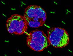
CHO cells dividing with isolated midbodies surrounding them. The cells and midbodies are stained with anti-actin (red), anti-tubulin (green) and DAPI (blue). This image shows Chinese hamster ovary cells in the last stages of division. The red outer membrane is complete around each new cell, while the green midbody still remains between them. Isolated midbodies are also pictured in green around the cells to show the organelles in more detail. <br>Photo by: courtesy Ahna Skop
Discovery may lead to insights into cancer, birth defects, fertility and neurological disorders
A cellular structure discovered 125 years ago and dismissed by many biologists as “cellular garbage” has been found to play a key role in the process of cytokinesis, or cell division, one of the most ancient and important of all biological phenomena.
The discovery of the function of the dozens of proteins harbored within this structure – which are necessary for normal cell division – by a team of scientists led by a University of Wisconsin-Madison geneticist was announced in today’s edition of the journal Science.
The discovery promises a better understanding of the role of cell division in the growth and development of all organisms and, critically, of abnormal cell division, when the key proteins fail. These failures can lead to infertility, birth defects, cancer and neurological problems such as Huntington’s and Alzheimer’s diseases.
“Going from one cell to two, or cytokinesis, is one of the most fundamental of cellular events,” dating to a time when life evolved from single-celled organisms, explains Ahna Skop, an assistant professor of genetics with the UW-Madison College of Agricultural and Life Sciences. “It applies to all species and organisms, and it is fundamental to the growth and development of all life on this planet.”
However, just as cell division is the key to life, failures in the process can lead to certain diseases, says Skop.
“Several diseases are caused by cells that don’t divide properly, or divide out of control, as in cancer,” she says. “In addition, proteins that work during cell division may also work in the neurons in our brain or during wound healing, for example. So understanding how cell division works can help us understand how many other specific types of cells function.”
With a new understanding of which proteins affect cell division, medical researchers can potentially develop new drugs to prevent cancer and birth defects, treat fertility and neurological disorders, or aide in wound healing, for example.
During the cell cycle, the genetic information the cell contains is copied and segregated into two new cells. During normal division, the outer membrane of the cell pinches in, forming two separate cells. Although a very simple event, scientists had not fully understood the mechanisms or identified the proteins involved in separating the two newly formed cells, says Skop.
As a graduate student at UW-Madison in John White’s laboratory, Skop became interested in an ephemeral cellular feature called the midbody, which forms briefly – lasting only a minute in the cells of some species – during cell division.
“It was identified over 125 years ago by Walther Flemming, but hadn’t been studied much or paid attention to since,” she explains. “Most people thought it was cellular garbage.”
However, Skop suspected that the midbody was more than an ancient relic with no useful function. She and colleagues used methods developed in the 1980s by Ryoko Kuriyama, then a UW-Madison postdoctoral researcher and now at the University of Minnesota, and Michael Mullins, now at Catholic University, to isolate midbodies from hamster ovary cells. They then analyzed and identified more than 500 proteins contained in the midbodies.
“Proteins are the building blocks of the cell,” Skop says. “As the cell divides and a new membrane forms, the proteins found during that time in the cell cycle would be crucial elements in understanding how the process works.”
The next step was to inactivate each protein in a developing embryo. If a defect occurred, it would mean that the inactivated protein is essential for normal development.
“We used nematodes, which are small roundworms that are cheaper and quicker to use than mammalian cells, to assess gene function,” Skop says. “All but two of the proteins from the mammalian cells were homologous in the nematodes, which allowed us to perform this mutli-organismal approach. The fact that the process is highly conserved across two very different species shows how ancient and conserved the process of cell division is.”
The team analyzed 160 key proteins – including 103 not previously known to function in cell division – and found that 58 percent caused cytokinesis defects if they were inactivated.
“The problems ranged from cells where chromosomes failed to separate normally, leaving extra DNA in one of the new cells, as is seen in Down’s syndrome, for example, to cells in which the dividing membrane would begin to form normally and would suddenly retract before the cells could separate,” Skop says. “Many of the proteins caused a variety of cell division and division-related defects.”
Skop, who also has an appointment with the UW-Madison Medical School, conducted some of this work as a postdoctoral researcher at the University of California, Berkeley. Her co-authors on the paper are Hongbin Liu and John Yates from the Scripps Research Institute, Rebecca Heald from UC Berkeley, and Barbara Meyer from UC Berkeley and the Howard Hughes Medical Institute.
The National Institutes of Health and the state of Wisconsin funded Skop’s work.
Media Contact
More Information:
http://www.news.wisc.edu/All latest news from the category: Life Sciences and Chemistry
Articles and reports from the Life Sciences and chemistry area deal with applied and basic research into modern biology, chemistry and human medicine.
Valuable information can be found on a range of life sciences fields including bacteriology, biochemistry, bionics, bioinformatics, biophysics, biotechnology, genetics, geobotany, human biology, marine biology, microbiology, molecular biology, cellular biology, zoology, bioinorganic chemistry, microchemistry and environmental chemistry.
Newest articles

An Endless Loop: How Some Bacteria Evolve Along With the Seasons
The longest natural metagenome time series ever collected, with microbes, reveals a startling evolutionary pattern on repeat. A Microbial “Groundhog Year” in Lake Mendota Like Bill Murray in the movie…

Witness Groundbreaking Research on Achilles Tendon Recovery
Achilles tendon injuries are common but challenging to monitor during recovery due to the limitations of current imaging techniques. Researchers, led by Associate Professor Zeng Nan from the International Graduate…

Why Prevention Is Better Than Cure—A Novel Approach to Infectious Disease Outbreaks
Researchers have come up with a new way to identify more infectious variants of viruses or bacteria that start spreading in humans – including those causing flu, COVID, whooping cough…


