Advances in understanding of the complexity of living cells: Molecular motors, tubes and adhesives, when a physicist meets a bio

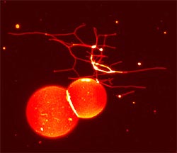
Molecular tubes to convey information 1. 2. Confocal microscopy of the network of membrane tubes. C. Leduc - P. Bassereau/Institut Curie
One of the Institut Curie’s great originalities, the interface between physics and cell biology, is a fertile terrain for discoveries. Dialogue between researchers of different backgrounds drives creativity, as witnessed by the rise in the number of Institut Curie publications on research work that melds physics and biology.
In collaboration with Canadian physicists, biologists of the (CNRS) group headed by Hélène Feracci have developed a model that cast light on intercellular adhesion. At the same time, the physicists of Patricia Bassereau’s (CNRS) team have worked with Institut Curie biologists and theoretical physicists to discover how communication works within cells. These studies were published in PNAS on 23 November and 7 December 2004, respectively.
Studies at the crossroads of physics and biology have their origins in the very creation of the Institut Curie, the brainchild of Marie Curie, Nobel Laureate in Physics and in Chemistry, and the oncologist Dr Claudius Régaud. The Institut Curie has since unfailingly encouraged interdisciplinary research and for many years now work at the physics-biology interface has been one of its most distinctive features. This work has now entered a new phase of maturity marked by an increase in publications.
Molecular adhesives to hold cells
In collaboration with Evan Evans and his team of physicists (Vancouver, Canada), Hélène Feracci, a biologist at the Institut Curie(1), has enhanced our understanding of the mechanisms of intercellular adhesion. United we stand, divided we fall: such could be the motto of epithelial cells. Organized in lamellae, epithelial cells cover outer surfaces, like the skin, or a cavity within the body, as the intestinal mucosa, and must remain attached to the tissue in order to function correctly. This cohesion is indispensable to correct functioning of the body and, like miniature soldiers, epithelial cells are expected to remain “dutifully” at their original tissue until they die.
Epithelial cells are held together by different molecular “adhesives”, including cadherins, surface glycoproteins which interact with themselves to maintain intercellular contacts. Cadherins play an essential part in tissue formation during embryonic development and in the cohesion of mature tissues. Perturbation of these interactions may have unfortunate consequences. When, for instance, cancer cells lose their ability to adhere to their neighbors, they may migrate and give rise to metastases.
To understand how these “adhesives” work, Hélène Feracci and Evan Evans utilized an experimental model: two glass beads coated with cadherins and a “spring” constituted by a tense red blood cell. The principle of this model is that a contact is established between two glass beads and the red cell “spring” is then used to measure the force needed to break this contact. Analysis of the spring force as a function of time provides information on the dynamic adhesive properties of cadherins.
This work shows that a single cadherin molecule has remarkable adhesive potential. Cadherins form contacts that are both dynamic, thus allowing them to adapt quickly to their environment, and highly stable, thereby favoring lasting interactions. This multidisciplinary approach casts new light on the complex mechanisms underpinning cellular adherence.
Molecular tubes to convey information
Intercellular communication, like its human counterpart, is essential, the very cornerstone of cellular existence. Exchanges between cells, and also between organelles within each cell, occur constantly and are vital to the maintenance of major biological functions.
To communicate, the cell uses molecules that “encode” information. Such molecules cannot, of course, be sent around the cell haphazardly – they need specialized carriers. Vital intracellular exchanges require the setting up of a genuine network to structure and prioritize information.
In partnership with the biologists of Bruno Goud’s (CNRS) team at the Institut Curie(2), the physicists in Patricia Bassereau’s (CNRS) group(3) have developed an original in vitro system which imitates intracellular traffic. The simplest system comprises lipid vesicles which constitute a reservoir of membrane and kinesins, and which act as molecular motors, with microtubules serving as rails and ATP as an energy source. In this way they have succeeded in forming very narrow tubes (a few dozen nanometers) similar to those seen in cells and which may carry the information(4).
Working with the group of theoretical physicists of Jacques Prost and Jean-François Joanny, they now know how the molecular motors – the kinesins – “tow” the tubes towards their destination: several motors, bound to the membrane and constantly renewed, combine dynamically to generate the force needed to pull the tubes. The towing speed is defined by the head of the motor.
The results seem to suggest that the cell can regulate its traffic by adjusting the concentration of kinesins or the vesicle membrane tension. The model developed by the Institut Curie physicists and biologists will continue to enhance our understanding of cellular traffic and signal transmission. Such understanding is overwhelmingly important since communication is essential to the organism and cancer can be regarded as a disease of signal transmission.
These studies perfectly illustrate the remarkable possibilities generated by cooperation between physicists and biologists. At the Institut Curie, and also worldwide, the physics-biology interface offers another vision of the world of the cell that is extremely promising for our attempts to penetrate the complexity of living systems.
(1)”Cellular morphogenesis and tumor progression” research team headed by Jean Paul Thiery in the UMR 144 CNRS/Institut Curie “Cellular compartmentalization and dynamics” group directed by Bruno Goud
(2) UMR 144 CNRS/Institut Curie “Cellular compartmentalization and dynamics” headed by Bruno Goud
(3) UMR 168 CNRS/Institut Curie “Physical Chemistry” headed by Jean-François Joanny
(4) “A minimal system allowing tubulation with molecular motors pulling on giant liposomes, A. Roux et al. PNAS, 16 April 2002, vol. 99, p. 5394-5399
Media Contact
All latest news from the category: Life Sciences and Chemistry
Articles and reports from the Life Sciences and chemistry area deal with applied and basic research into modern biology, chemistry and human medicine.
Valuable information can be found on a range of life sciences fields including bacteriology, biochemistry, bionics, bioinformatics, biophysics, biotechnology, genetics, geobotany, human biology, marine biology, microbiology, molecular biology, cellular biology, zoology, bioinorganic chemistry, microchemistry and environmental chemistry.
Newest articles

Parallel Paths: Understanding Malaria Resistance in Chimpanzees and Humans
The closest relatives of humans adapt genetically to habitats and infections Survival of the Fittest: Genetic Adaptations Uncovered in Chimpanzees Görlitz, 10.01.2025. Chimpanzees have genetic adaptations that help them survive…

You are What You Eat—Stanford Study Links Fiber to Anti-Cancer Gene Modulation
The Fiber Gap: A Growing Concern in American Diets Fiber is well known to be an important part of a healthy diet, yet less than 10% of Americans eat the minimum recommended…

Trust Your Gut—RNA-Protein Discovery for Better Immunity
HIRI researchers uncover control mechanisms of polysaccharide utilization in Bacteroides thetaiotaomicron. Researchers at the Helmholtz Institute for RNA-based Infection Research (HIRI) and the Julius-Maximilians-Universität (JMU) in Würzburg have identified a…


