Through the backdoor: How phosphate escapes from actin

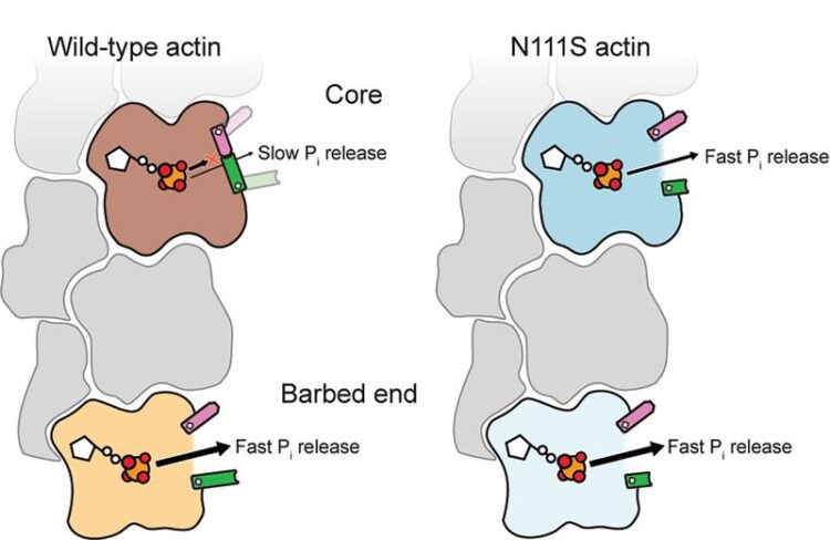
Model showing the release of phosphate in different parts of the actin filament. Actin in the core of the filament has a closed door while the back door at the end of the filament is open. A mutation opens the door in the core.
(c) MPI of Molecular Physiology
MPI scientists reveal how phosphate escapes from actin filaments – a key signal that primes older filaments for disassembly.
Actin filaments are dynamic protein-fibres in the cell built from single actin proteins. Many cellular functions, including cell movement, are regulated by constant filament assembly and disassembly. The disassembly phase is initiated by the release of a phosphate group from inside the filament, but the details of this process have puzzled scientists since decades. Researchers from the Max Planck Institute of Molecular Physiology in Dortmund and the Max Planck Institute of Biophysics in Frankfurt have joined forces to precisely identify a region in actin that functions as a “molecular backdoor” for phosphate to exit though. Using a wide variety of techniques, including cryogenic electron microscopy (cryo-EM) and molecular dynamics simulations, the scientists determined the mechanism of phosphate release from actin filaments in unprecedented molecular detail. They also described how a distorted backdoor enables the faster release of phosphate from an actin mutant linked to nemaline myopathy, a severe muscle disease. The study opens the door to further research on the dynamic actin-assembly cycle in cells and diseases related to defective actin organization.
The mysterious escape of phosphate
In eukaryotic cells, actin proteins join together (polymerize) into filaments that are part of the cell’s intricate supportive network, the cytoskeleton. The disassembly of old filaments is crucial for cell movement and is regulated by ATP hydrolysis – the reaction of ATP with water that cleaves a phosphate group and generates energy. Specifically, phosphate release from the filament core is the signal to the cell that the actin filament is old enough and can be dismantled into actin subunits. “The mechanism of phosphate release from actin filaments has remained enigmatic for decades,” says Wout Oosterheert, postdoc in the group of Stefan Raunser at the MPI Dortmund and first author of the publication.

Phosphate escapes through molecular backdoor in actin. (c) MPI of Molecular Physiology
The new results are built on previous research of Raunser’s group on actin that led to ground-breaking publications in 2015, 2018, and 2022 in the actin field. In the latter, the Raunser team determined high-resolution cryo-EM structures of actin filaments in three different states: bound to ATP, bound to ADP in the presence of the cleaved phosphate, and bound to ADP after release of the phosphate. However, in all structures, there was no opening or door in actin through which phosphate could escape from the filament. “Therefore, we surmised that there should be a backdoor that opens momentarily to release the phosphate, and then quickly closes again” says Raunser.
A multidisciplinary approach
MPI scientists have now tackled the problem from various angles. Since it was known that phosphate is released very rapidly from actin at the tip of the filament, called the barbed end, Raunser and his team determined its structure by cryo-EM. And indeed, only at the end of the filament, they found an open molecular backdoor, which explains the very fast phosphate release. However, it was still unclear how phosphate escapes from the actin subunits in the filament core. That’s where the expertise of Gerhard Hummer’s group from the MPI Frankfurt kicked in; they used the structural data from 2022 to perform molecular dynamics simulations and predict potential exit routes for the phosphate from the filament core. They then teamed up with the group of Peter Bieling (MPI Dortmund) to validate the possible routes by producing actin mutants potentially disrupting the molecular backdoor. They measured how fast they release the phosphate, and finally determined the high-resolution cryo-EM structures of the “fastest” candidates.
The mutational analysis revealed that the phosphate takes the same release route in the filament end and the filament core. The structures and interactions in the latter, however, need additional rearrangements that make it more difficult for the door to open. After phosphate cleavage, the backdoor remains predominantly closed (on average for 100 seconds) before opening for less than a second to let the phosphate leave. “This explains why we didn’t see an open backdoor arrangement in our cryo-EM data of 2022,” says Raunser.
The actin saga – To be continued…
One of the actin mutants analyzed, called N111S, is linked to the muscular disease nemaline myopathy and has therefore attracted the attention of the MPI scientists: the mutant always adopts an open backdoor and hence releases phosphate much faster than normal wild-type actin. “We propose that this ultrafast release may contribute to the pathophysiology in patients harboring this actin mutation,” says Oosterheert.
As a potential next step, the MPI scientists now want to uncover how phosphate release is controlled within the cell and what role the proteins that bind to actin play. In addition, their work now makes it possible to investigate other disease-related mutations in actin – an approach that may ultimately contribute to the development of new therapeutic strategies for these diseases.
Original publication:
Oosterheert W, Blanc FEC, Roy A, Belyy A, Sanders MB, Hofnagel O, Hummer G, Bieling P, Raunser S (2023). Molecular mechanisms of inorganic-phosphate release from the core and barbed end of actin filaments. Nat Struct Mol Biol. Doi: 10.1038/s41594-023-01101-9.
More information:
https://www.mpi-dortmund.mpg.de/news/actin-backdoor-phosphate
Media Contact
All latest news from the category: Life Sciences and Chemistry
Articles and reports from the Life Sciences and chemistry area deal with applied and basic research into modern biology, chemistry and human medicine.
Valuable information can be found on a range of life sciences fields including bacteriology, biochemistry, bionics, bioinformatics, biophysics, biotechnology, genetics, geobotany, human biology, marine biology, microbiology, molecular biology, cellular biology, zoology, bioinorganic chemistry, microchemistry and environmental chemistry.
Newest articles

Innovative 3D printed scaffolds offer new hope for bone healing
Researchers at the Institute for Bioengineering of Catalonia have developed novel 3D printed PLA-CaP scaffolds that promote blood vessel formation, ensuring better healing and regeneration of bone tissue. Bone is…

The surprising role of gut infection in Alzheimer’s disease
ASU- and Banner Alzheimer’s Institute-led study implicates link between a common virus and the disease, which travels from the gut to the brain and may be a target for antiviral…

Molecular gardening: New enzymes discovered for protein modification pruning
How deubiquitinases USP53 and USP54 cleave long polyubiquitin chains and how the former is linked to liver disease in children. Deubiquitinases (DUBs) are enzymes used by cells to trim protein…


