What you count is not necessarily what counts

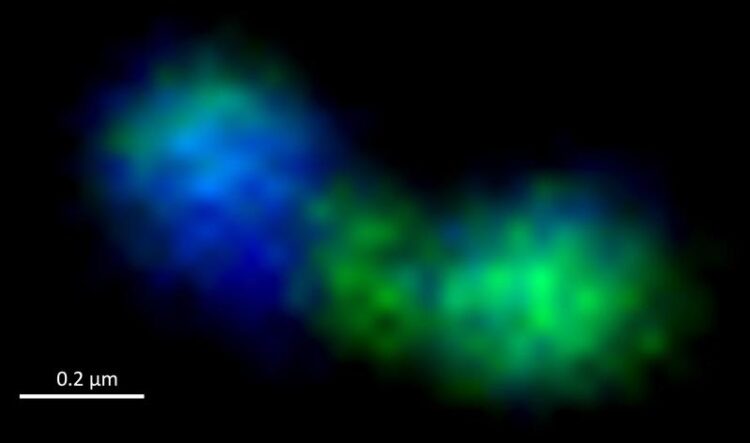
A dividing bacterial cell under the fluorescence microscope. The constriction in the middle where the cell divides is clearly visible. The cell is coloured green, the genomes blue.
(c) Jan Brüwer / Max Planck Institute for Marine Microbiology
A new study focusing on the number of dividing bacterial cells in the North Sea challenges some dogmas about marine microbial life.
Seawater is full of bacteria, hundreds of thousands live in every litre. But the sheer number of bacteria living in the water does not necessarily mean a lot. More important is how active they are and how quickly they duplicate, as demonstrated in a study by researchers from the Max Planck Institute for Marine Microbiology in Bremen, now published in the journal mSystems.
If scientists want to find out how fast a population of bacteria grows, they often measure how their cell count changes over time. However, this method has a major flaw: it does not measure how fast the bacteria multiply or die. Yet these factors are very important for understanding ecological processes. That is why researchers at the Max Planck Institute for Marine Microbiology in Bremen have now taken a closer look at these processes during a spring bloom in the German Bight. In doing so, they challenge some previous dogmas.
The researchers around Jan Brüwer, Bernhard Fuchs and Rudolf Amann investigated the growth of bacteria during the spring bloom off Helgoland using various methods: With the microscope, they counted and identified not only the cells present, but also the frequency of cells that were currently dividing. This way, they were able to calculate how quickly different types of bacteria multiplied in their natural environment.
“We used modern microscopic methods to visualise and count dividing cells in thousands of images,” explains Jan Brüwer, who conducted the study as part of his doctoral thesis. “We made use of the fact that a dividing cell has to split its duplicated genome into its daughter cells. Thus, we were able to clearly identify these cells based on the DNA distribution in the cell.” This enabled the researchers to determine the growth rates of individual groups of bacteria over longer periods of time.
“The results had some surprises in store for us,” says group leader Bernhard Fuchs. “For example, we found that the most common group of bacteria in the ocean, called SAR11, divides almost ten times faster than assumed.” Moreover, in many cases the measured growth rates do not match the abundance of the respective bacteria in the water. “If bacteria divide often but are not abundant, it suggests that they are a popular victim of predators or viruses,” Brüwer explains. “The timing of bacterial proliferation was also surprising: SAR11 bacteria frequently divided before the onset of the algal bloom in the North Sea. From where they took the required energy to do so is still a mystery.”
Not all bacterial groups behaved as unexpectedly as SAR11; for other groups, the results now collected were more in line with the researchers’ expectations – in their case, growth rates and cell numbers largely matched.
Until now, it has been assumed that SAR11, which have very small cells, get by with little nutrients, do not divide very often and are eaten only rarely because of their small size. In contrast, other larger bacteria, for example the Bacteroidetes, are seen as popular food, multiplying quickly and disappearing just as quickly when predators and viruses get on their trail. The new study by Brüwer and his colleagues paints a very different picture.
“Our results influence our understanding of element cycles, especially the carbon cycle, in the ocean,” Brüwer emphasises. “The most abundant bacteria in the ocean, SAR11, are more active and divide faster than previously believed. This could mean that they need fewer nutrients and are a more popular food source for other organisms than suspected. Also, the general turnover of bacteria during algal blooms seems to be faster than we thought.”
“This research was methodologically very demanding and it shows how much information you can draw from microscopy images,” stresses Rudolf Amann, director at the Max Planck Institute for Marine Microbiology. “I am very proud of the researchers involved for mastering this mammoth task and glad to have the privilege to work with them. The results achieved will trigger many exciting discussions about the ecological relationships during a spring bloom and in the ocean in general.”
Wissenschaftliche Ansprechpartner:
Jan Brüwer
Department of Molecular Ecology, Flow Cytometry Research Group
Max Planck Institute for Marine Microbiology, Bremen, Germany
Phone: +49 421 2028-9280
E-mail: jbruewer@mpi-bremen.de
PD Dr. Bernhard Fuchs
Department of Molecular Ecology, Flow Cytometry Research Group
Max Planck Institute for Marine Microbiology, Bremen, Germany
Phone: +49 421 2028-9350
E-mail: bfuchs@mpi-bremen.de
Dr. Fanni Aspetsberger
Press Officer
Max Planck Institute for Marine Microbiology, Bremen, Germany
Phone: +49 421 2028-9470
E-mail: presse@mpi-bremen.de
Originalpublikation:
Brüwer, JD, Orellana, LH, Sidhu, C, Klip, HCL, Meunier, CL, Boersma, M, Wiltshire, KH, Amann, R, Fuchs, BM (2023): In situ cell division and mortality rates of SAR11, SAR86, Bacteroidetes, and Aurantivirga during phytoplankton blooms reveal differences in population controls. mSystems (17 May 2023)
DOI: 10.1128/msystems.01287-22
Weitere Informationen:
Media Contact
All latest news from the category: Life Sciences and Chemistry
Articles and reports from the Life Sciences and chemistry area deal with applied and basic research into modern biology, chemistry and human medicine.
Valuable information can be found on a range of life sciences fields including bacteriology, biochemistry, bionics, bioinformatics, biophysics, biotechnology, genetics, geobotany, human biology, marine biology, microbiology, molecular biology, cellular biology, zoology, bioinorganic chemistry, microchemistry and environmental chemistry.
Newest articles

First-of-its-kind study uses remote sensing to monitor plastic debris in rivers and lakes
Remote sensing creates a cost-effective solution to monitoring plastic pollution. A first-of-its-kind study from researchers at the University of Minnesota Twin Cities shows how remote sensing can help monitor and…

Laser-based artificial neuron mimics nerve cell functions at lightning speed
With a processing speed a billion times faster than nature, chip-based laser neuron could help advance AI tasks such as pattern recognition and sequence prediction. Researchers have developed a laser-based…

Optimising the processing of plastic waste
Just one look in the yellow bin reveals a colourful jumble of different types of plastic. However, the purer and more uniform plastic waste is, the easier it is to…


