A new brain-computer interface with a flexible backing

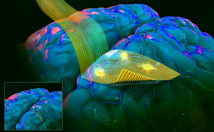
Artist rendition of the flexible, conformable, transparent backing of the new brain-computer interface with penetrating microneedles developed by a team led by engineers at the University of California San Diego in the laboratory of electrical engineering professor Shadi Dayeh. The smaller illustration at bottom left shows the current technology in experimental use called Utah Arrays.
Credit: Shadi Dayeh / UC San Diego / SayoStudio
The flexible backing allows arrays of micro-scale needles to conform to the contours of the brain, which improves high-resolution brain recording.
Engineering researchers have invented an advanced brain-computer interface with a flexible and moldable backing and penetrating microneedles. Adding a flexible backing to this kind of brain-computer interface allows the device to more evenly conform to the brain’s complex curved surface and to more uniformly distribute the microneedles that pierce the cortex. The microneedles, which are 10 times thinner than the human hair, protrude from the flexible backing, penetrate the surface of the brain tissue without piercing surface venules, and record signals from nearby nerve cells evenly across a wide area of the cortex.
This novel brain-computer interface has thus far been tested in rodents. The details were published online on February 25 in the journal Advanced Functional Materials. This work is led by a team in the lab of electrical engineering professor Shadi Dayeh at the University of California San Diego, together with researchers at Boston University led by biomedical engineering professor Anna Devor.
This new brain-computer interface is on par with and outperforms the “Utah Array,” which is the existing gold standard for brain-computer interfaces with penetrating microneedles. The Utah Array has been demonstrated to help stroke victims and people with spinal cord injury. People with implanted Utah Arrays are able to use their thoughts to control robotic limbs and other devices in order to restore some everyday activities such as moving objects.
The backing of the new brain-computer interface is flexible, conformable, and reconfigurable, while the Utah Array has a hard and inflexible backing. The flexibility and conformability of the backing of the novel microneedle-array favors closer contact between the brain and the electrodes, which allows for better and more uniform recording of the brain-activity signals. Working with rodents as model species, the researchers have demonstrated stable broadband recordings producing robust signals for the duration of the implant which lasted 196 days.
In addition, the way the soft-backed brain-computer interfaces are manufactured allows for larger sensing surfaces, which means that a significantly larger area of the brain surface can be monitored simultaneously. In the Advanced Functional Materials paper, the researchers demonstrate that a penetrating microneedle array with 1,024 microneedles successfully recorded signals triggered by precise stimuli from the brains of rats. This represents ten times more microneedles and ten times the area of brain coverage, compared to current technologies.
Thinner and transparent backings
These soft-backed brain-computer interfaces are thinner and lighter than the traditional, glass backings of these kinds of brain-computer interfaces. The researchers note in their Advanced Functional Materials paper that light, flexible backings may reduce irritation of the brain tissue that contacts the arrays of sensors.
The flexible backings are also transparent. In the new paper, the researchers demonstrate that this transparency can be leveraged to perform fundamental neuroscience research involving animal models that would not be possible otherwise. The team, for example, demonstrated simultaneous electrical recording from arrays of penetrating micro-needles as well as optogenetic photostimulation.
Two-sided lithographic manufacturing
The flexibility, larger microneedle array footprints, reconfigurability and transparency of the backings of the new brain sensors are all thanks to the double-sided lithography approach the researchers used.
Conceptually, starting from a rigid silicon wafer, the team’s manufacturing process allows them to build microscopic circuits and devices on both sides of the rigid silicon wafer. On one side, a flexible, transparent film is added on top of the silicon wafer. Within this film, a bilayer of titanium and gold traces is embedded so that the traces line up with where the needles will be manufactured on the other side of the silicon wafer.
Working from the other side, after the flexible film has been added, all the silicon is etched away, except for free-standing, thin, pointed columns of silicon. These pointed columns of silicon are, in fact, the microneedles, and their bases align with the titanium-gold traces within the flexible layer that remains after the silicon has been etched away. These titanium-gold traces are patterned via standard and scalable microfabrication techniques, allowing scalable production with minimal manual labor. The manufacturing process offers the possibility of flexible array design and scalability to tens of thousands of microneedles.
Toward closed-loop systems
Looking to the future, penetrating microneedle arrays with large spatial coverage will be needed to improve brain-machine interfaces to the point that they can be used in “closed-loop systems” that can help individuals with severely limited mobility. For example, this kind of closed-loop system might offer a person using a robotic hand real-time tactical feedback on the objects the robotic hand is grasping.
Tactile sensors on the robotic hand would sense the hardness, texture, and weight of an object. This information recorded by the sensors would be translated into electrical stimulation patterns which travel through wires outside the body to the brain-computer interface with penetrating microneedles. These electrical signals would provide information directly to the person’s brain about the hardness, texture, and weight of the object. In turn, the person would adjust their grasp strength based on sensed information directly from the robotic arm.
This is just one example of the kind of closed-loop system that could be possible once penetrating microneedle arrays can be made larger to conform to the brain and coordinate activity across the “command” and “feedback” centers of the brain.
Previously, the Dayeh laboratory invented and demonstrated the kinds of tactile sensors that would be needed for this kind of application, as highlighted in this video.
Pathway to commercialization
The advanced dual-side lithographic microfabrication processes described in this paper are patented (US 10856764). Dayeh co-founded Precision Neurotek Inc. to translate technologies innovated in his laboratory to advance state of the art in clinical practice and to advance the fields of neuroscience and neurophysiology.
Paper title: “Scalable Thousand Channel Penetrating Microneedle Arrays on Flex for Multimodal and Large Area Coverage BrainMachine Interfaces,” in Advanced Functional Materials
Authors
S. H. Lee, K. Lee, D. R. Cleary, K. J. Tonsfeldt, H. Oh, F. Azzazy, Y. Tchoe, A. M. Bourhis, L. Hossain, Y. G. Ro, A. Tanaka, S. A. Dayeh are affiliated with the Integrated Electronics and Biointerfaces Laboratory in the Department of Electrical and Computer Engineering at the University of California San Diego Jacobs School of Engineering.
D. R. Cleary has a primary appointment in the Department of Neurological Surgery at UC San Diego Health.
M. Thunemann, K. Kılıç, and A. Devor are affiliated with the Biomedical Engineering Department at Boston University.
Corresponding author
Shadi Dayeh
Electrical and Computer Engineering Department
UC San Diego Jacobs School of Engineering
Email: sdayeh@eng.ucsd.edu
Funding
This work was supported by the National Institutes of Health Awards NIBIB DP2-EB029757, the NIH BRAIN Initiative R01NS123655-01,UG3NS123723-01 to S.A.D., 1R01DA050159-01, R01 MH111359-05 to A.D., and F32 MH120886-01 to D.R.C.; National Science Foundation Award No. 1728497 and CAREER No. 1351980; and by the KAVLI Institute for Brain and Mind to S.A.D.. Technical support from the Nano3 cleanroom facilities at UC San Diego’s Qualcomm Institute. This work was performed in part at the San Diego Nanotechnology Infrastructure (SDNI) of UC San Diego, a member of the National Nanotechnology Coordinated Infrastructure, which was supported by the National Science Foundation (Grant No. ECCS1542148).
Journal: Advanced Functional Materials
DOI: 10.1002/adfm.202112045
Method of Research: Experimental study
Subject of Research: Animals
Article Title: Scalable Thousand Channel Penetrating Microneedle Arrays on Flex for Multimodal and Large Area Coverage BrainMachine Interfaces
Article Publication Date: 25-Feb-2022
COI Statement: The authors declare no conflict of interest.
Media Contact
Daniel Kane
University of California – San Diego
dbkane@ucsd.edu
Office: 858-534-3262
Original Source
All latest news from the category: Medical Engineering
The development of medical equipment, products and technical procedures is characterized by high research and development costs in a variety of fields related to the study of human medicine.
innovations-report provides informative and stimulating reports and articles on topics ranging from imaging processes, cell and tissue techniques, optical techniques, implants, orthopedic aids, clinical and medical office equipment, dialysis systems and x-ray/radiation monitoring devices to endoscopy, ultrasound, surgical techniques, and dental materials.
Newest articles

Innovative 3D printed scaffolds offer new hope for bone healing
Researchers at the Institute for Bioengineering of Catalonia have developed novel 3D printed PLA-CaP scaffolds that promote blood vessel formation, ensuring better healing and regeneration of bone tissue. Bone is…

The surprising role of gut infection in Alzheimer’s disease
ASU- and Banner Alzheimer’s Institute-led study implicates link between a common virus and the disease, which travels from the gut to the brain and may be a target for antiviral…

Molecular gardening: New enzymes discovered for protein modification pruning
How deubiquitinases USP53 and USP54 cleave long polyubiquitin chains and how the former is linked to liver disease in children. Deubiquitinases (DUBs) are enzymes used by cells to trim protein…


