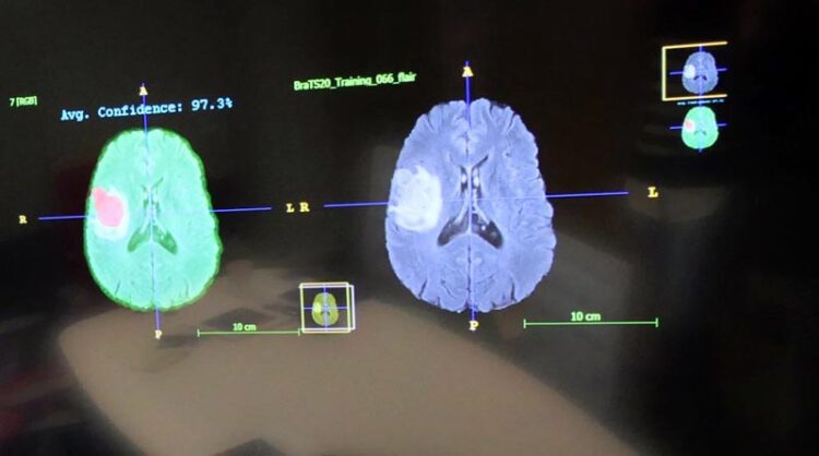
Evaluating Brain Tumours with Artificial Intelligence

The evaluation of tumours in MRI images is an important diagnostic step that can be supported by AI.
Picture: TU Darmstadt
Best Paper Award: Outstanding Publication by TU Researchers Recognised.
One application area of artificial intelligence (AI) is in medicine, especially in medical diagnostics. For instance, scans can be analysed automatically with the help of algorithms. An international and interdisciplinary team led by researchers from TU Darmstadt recently investigated whether AI can better evaluate images of brain tumours. For this publication, the team won the Best Paper Award at the world’s largest information systems conference ICIS, prevailing over more than 1,300 other publications.
The information systems department of TU Darmstadt regularly achieves very good rankings, such as second place in the university ranking of the magazine “WirtschaftsWoche”. The research results are also impressive. An international team of researchers from TU Darmstadt, the University of Cambridge, the science and technology company Merck, and the Klinikum rechts der Isar of TU Munich, studied in an international and interdisciplinary collaboration how software systems collect, process, and evaluate task-specific relevant information, thereby supporting the work of humans, in this case, radiologists. The study, now awarded with the Best Paper Award, provides empirical data on the influence of machine learning systems (ML systems) on human learning. It also shows how important it is for end users whether the results of machine learning methods are comprehensible and understandable. These insights are not only relevant for medical diagnoses in radiology, but for everyone who becomes a reviewer of ML output through the daily use of AI tools, such as ChatGPT.
The research project, led by TU researchers Sara Ellenrieder and Professor Peter Buxmann, investigated the use of ML-based decision support systems in radiology, specifically in the manual segmentation of brain tumours in MRI images. The focus was on how radiologists can learn from these systems to improve their performance and decision-making confidence. The authors compared different performance levels of ML systems and analyzed how explaining the ML output improved the radiologists’ understanding of the results. The research aim is to find out how radiologists can benefit from these systems in the long term and use them safely.
For this purpose, the project team conducted an experiment with radiologists from various clinics. The physicians were asked to segment tumours in MRI images before and after receiving ML-based decision support. Different groups were provided with ML systems of varying performance or explainability. In addition to collecting quantitative performance data during the experiment, the researchers also gathered qualitative data through “think-aloud” protocols and subsequent interviews.
In the experiment, 690 manual segmentations of brain tumours were performed by the radiologists.
The results show that radiologists can learn from the information provided by high-performing ML systems. Through interaction, they improved their performance. However, the study also shows that a lack of explainability of ML output in low-performing systems can lead to a decline in performance among radiologists. Interestingly, providing explanations of the ML output not only improved the learning outcomes of the radiologists but also prevented learning false information. In fact, some physicians were even able to learn from mistakes made by low-performing, but explainable systems.
“The future of human-AI collaboration lies in the development of explainable and transparent AI systems that enable end users in particular to learn from the systems and make better decisions in the long term,” summarises Professor Peter Buxmann from TU Darmstadt.
About TU Darmstadt
TU Darmstadt is one of Germany’s leading technical universities and a synonym for excellent, relevant research. We are crucially shaping global transformations – from the energy transition via Industry 4.0 to artificial intelligence – with outstanding insights and forward-looking study opportunities.
TU Darmstadt pools its cutting-edge research in three fields: Energy and Environment, Information and Intelligence, Matter and Materials. Our problem-based interdisciplinarity as well as our productive interaction with society, business and politics generate progress towards sustainable development worldwide.
Since we were founded in 1877, we have been one of Germany’s most international universities; as a European technical university, we are developing a trans-European campus in the network, Unite! With our partners in the alliance of Rhine-Main universities – Goethe University Frankfurt and Johannes Gutenberg University Mainz – we further the development of the metropolitan region Frankfurt-Rhine-Main as a globally attractive science location.
MI-Nr. 50e/2023, cst/mho
Wissenschaftliche Ansprechpartner:
Prof. Dr. Peter Buxmann
E-Mail: Peter.buxmann@tu-darmstadt.de
Sara Ellenrieder M.Sc.
E-Mail: sara.ellenrieder@tu-darmstadt.de
Originalpublikation:
Sara Ellenrieder, Emma Marlene Kallina, Luisa Pumplun, Joshua Felix Gawlitza, Sebastian Ziegelmayer, Peter Buxmann: Promoting Learning Through Explainable Artificial Intelligence: An Experimental Study in Radiology
https://www.tu-darmstadt.de/universitaet/aktuelles_meldungen/einzelansicht_433088.en.jsp
Media Contact
All latest news from the category: Medical Engineering
The development of medical equipment, products and technical procedures is characterized by high research and development costs in a variety of fields related to the study of human medicine.
innovations-report provides informative and stimulating reports and articles on topics ranging from imaging processes, cell and tissue techniques, optical techniques, implants, orthopedic aids, clinical and medical office equipment, dialysis systems and x-ray/radiation monitoring devices to endoscopy, ultrasound, surgical techniques, and dental materials.
Newest articles

Largest magnetic anisotropy of a molecule measured at BESSY II
At the Berlin synchrotron radiation source BESSY II, the largest magnetic anisotropy of a single molecule ever measured experimentally has been determined. The larger this anisotropy is, the better a…

Breaking boundaries: Researchers isolate quantum coherence in classical light systems
LSU quantum researchers uncover hidden quantum behaviors within classical light, which could make quantum technologies robust. Understanding the boundary between classical and quantum physics has long been a central question…

MRI-first strategy for prostate cancer detection proves to be safe
Active monitoring is a sufficiently safe option when prostate MRI findings are negative. There are several strategies for the early detection of prostate cancer. The first step is often a…


