Ga-68 FAPI PET improves detection and staging of pancreatic cancer

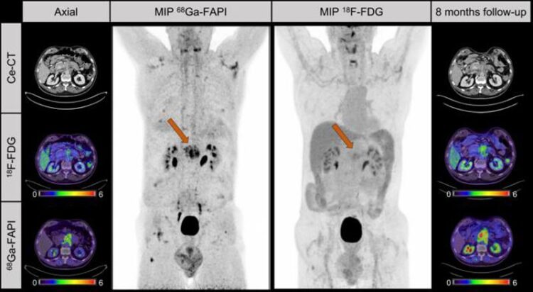
Figure 3. Case presentation. Male patient with suspected recurrent pancreatic cancer. Ce-CT shows mass around superior mesenteric artery after pancreatectomy. 18F-FDG only shows discrete uptake of lesion (SUVmax, 3.8), whereas 68Ga-FAPI clearly visualizes recurrent tumor. Patient received adjuvant chemotherapy after 68Ga-FAPI. Patient denied recommended chemotherapy. Increase of CA19-9 levels (from 240 to 767 U/mL) and follow-up imaging 8mo later validated progression of mesenteric mass as well as increased 18F-FDG uptake (SUVmax, 5.3). MIP 5 maximum-intensity projection.
Image created by L. Kessler, MD, University Hospital Essen, Essen, Germany
PET imaging with 68Ga-FAPI can more effectively detect and stage pancreatic cancer as compared with 18F-FDG imaging or contrast-enhanced CT, according to new research published in the December issue of The Journal of Nuclear Medicine. In a head-to-head study, 68Ga-FAPI detected more pancreatic tumors on a per-lesion, per-patient, or per-region basis and led to major and minor changes to clinical management of patients. In addition to enhancing precise detection of pancreatic cancer, 68Ga-FAPI imaging also paves the way for future targeted radiopharmaceutical therapies.
Approximately 64,000 Americans are diagnosed with pancreatic cancer each year. The disease is often diagnosed in advanced or metastasized stages and, as a result, is associated with extremely poor survival.
“Existing diagnostic approaches and workups are not sufficient for early detection of pancreatic cancer in curative stages for most patients,” said Jens T. Siveke, MD, translational and GI oncologist of the German Cancer Consortium (DKTK) at the West German Cancer Center in Essen, Germany. “Consequently, there is a pressing need for earlier and more precise disease detection, as well as a demand for novel targeted therapies.”
Recent studies have demonstrated high radiotracer uptake of 68Ga-FAPI in pancreatic cancer lesions; however, the precise diagnostic accuracy and the correlation of the tracer remain unexplored. In this study researchers sought to provide comprehensive data on the diagnostic performance of 68Ga-FAPI in pancreatic cancer patients.
Sixty-four patients with suspected or proven pancreatic cancer were included in the study. All patients underwent 68Ga-FAPI PET and contrast-enhanced CT, and 38 of the patients also underwent 18F-FDG PET. Researchers observed the association of the 68Ga-FAPI PET uptake intensity and histologic FAP (fibroblast activation protein) expression. The detection rate, diagnostic performance, inter-reader reproducibility, and change in management were also analyzed.
The association between 68Ga-FAPI PET uptake intensity and FAP expression was found to be significant, and 68Ga-FAPI PET showed high sensitivity and positive predictive values. In a head-to-head comparison with 18F-FDG and contrast-enhanced CE, 68Ga-FAPI PET detected more tumors on a per-lesion (84.7 vs. 46.5 vs. 52.9 percent), per-patient (97.4 vs. 73.7 vs. 92.1 percent), or per-region (32.6 vs. 18.8 vs. 23.7 percent) basis, respectively. 68Ga-FAPI PET readers showed substantial overall agreement, and minor and major changes in clinical management occurred in nearly 10 percent of patients after 68Ga-FAPI PET.
“Our research suggests that 68Ga-FAPI could become a building block in the diagnostic work-up of pancreatic cancer to improve early detection and accurate staging of this disease,” noted Lukas Kessler, MD, resident in the Department of Nuclear Medicine at University Hospital Essen in Germany. “Furthermore, our results support further investigation of FAP as a potential theranostic target of the tumor microenvironment, which represents an exciting new avenue in combating this enigmatic and fatal disease.”
This study was made available online in November 2023.
The authors of “68Ga-Labeled Fibroblast Activation Protein Inhibitor (68Ga-FAPI) PET for Pancreatic Adenocarcinoma: Data from the 68Ga-FAPI PET Observational Trial” include Lukas Kessler, Department of Nuclear Medicine, University Hospital Essen, University of Duisburg-Essen, Essen, Germany, Department of Diagnostic and Interventional Radiology and Neuroradiology, University Hospital Essen, Essen, Germany, and German Cancer Consortium (DKTK) (Partner Site University Hospital Essen) and German Cancer Research Center (DKFZ), Essen, Germany; Nader Hirmas, Ken Herrmann, and Wolfgang P. Fendler, Department of Nuclear Medicine, University Hospital Essen, University of Duisburg-Essen, Essen, Germany, and German Cancer Consortium (DKTK) (Partner Site University Hospital Essen) and German Cancer Research Center (DKFZ), Essen, Germany; Kim M. Pabst and Justin Ferdinandus, Department of Nuclear Medicine, University Hospital Essen, University of Duisburg-Essen, Essen, Germany, and Department of Diagnostic and Interventional Radiology and Neuroradiology, University Hospital Essen, Essen, Germany; Rainer Hamacher, German Cancer Consortium (DKTK) (Partner Site University Hospital Essen) and German Cancer Research Center (DKFZ), Essen, Germany, and Department of Medical Oncology, West German Cancer Center, University of Duisburg-Essen, Essen, Germany; Benedikt M. Schaarschmidt, Aleksander Milosevic, and Lale Umutlu, Department of Diagnostic and Interventional Radiology and Neuroradiology, University Hospital Essen, Essen, Germany, and German Cancer Consortium (DKTK) (Partner Site University Hospital Essen) and German Cancer Research Center (DKFZ), Essen, Germany; Michael Nader, Department of Nuclear Medicine, University Hospital Essen, University of Duisburg-Essen, Essen, Germany; Waldemar Uhl, Department of General and Visceral Surgery, St. Josef Hospital Bochum, Ruhr-University Bochum, Bochum, Germany; Anke Reinacher-Schick, Department of Hematology and Oncology with Palliative Care, St. Josef-Hospital, Ruhr-University Bochum, Bochum, Germany; Celine Lugnier, David Witter, and Marco Niedergethmann, Department of General and Visceral Surgery, Alfried Krupp Hospital, Essen, Germany; and Jens T. Siveke, German Cancer Consortium (DKTK) (Partner Site University Hospital Essen) and German Cancer Research Center (DKFZ), Essen, Germany, Bridge Institute of Experimental Tumor Therapy, West German Cancer Center, University Hospital Essen, University of Duisburg-Essen, Essen, Germany, and Division of Solid Tumor Translational Oncology, German Cancer Consortium (DKTK) (Partner Site University Hospital Essen) and German Cancer Research Center (DKFZ), Heidelberg, Germany.
Visit the JNM website for the latest research, and follow our new Twitter and Facebook pages @JournalofNucMed or follow us on LinkedIn.
Please visit the SNMMI Media Center for more information about molecular imaging and precision imaging. To schedule an interview with the researchers, please contact Rebecca Maxey at (703) 652-6772 or rmaxey@snmmi.org.
About JNM and the Society of Nuclear Medicine and Molecular Imaging
The Journal of Nuclear Medicine (JNM) is the world’s leading nuclear medicine, molecular imaging and theranostics journal, accessed 15 million times each year by practitioners around the globe, providing them with the information they need to advance this rapidly expanding field. Current and past issues of The Journal of Nuclear Medicine can be found online at http://jnm.snmjournals.org.
JNM is published by the Society of Nuclear Medicine and Molecular Imaging (SNMMI), an international scientific and medical organization dedicated to advancing nuclear medicine and molecular imaging—precision medicine that allows diagnosis and treatment to be tailored to individual patients in order to achieve the best possible outcomes. For more information, visit www.snmmi.org.
Journal: Journal of Nuclear Medicine
DOI: 10.2967/jnumed.122.264827
Article Title: 68Ga-Labeled Fibroblast Activation Protein Inhibitor (68Ga-FAPI) PET for Pancreatic Adenocarcinoma: Data from the 68Ga-FAPI PET Observational Trial
Article Publication Date: 1-Dec-2023
COI Statement: Ken Herrmann and Jens T. Siveke are supported by the German Federal Ministry of Education and Research (BMBF; 01KD2206A/SATURN3). The work of Jens T. Siveke is supported by the Deutsche Forschungsgemeinschaft (DFG, German Research Foundation) through 405344257 (SI1549/3-2) and SI1549/4-1, by the German Cancer Consortium (DKTK), and by German Cancer Aid (70112505/PIPAC, 70113834/PREDICT-PACA). Lukas Kessler is consultant for BTG and AAA and has received fees from Sanofi outside of the submitted work. Kim M. Pabst has received a Junior Clinician Scientist Stipend granted by the University Duisburg-Essen, travel fees from IPSEN, and research funding from Bayer. Rainer Hamacher has received travel grants from Lilly, Novartis, and PharmaMar as well as fees from Lilly outside of the submitted work and is supported by the Clinician Scientist Program of the University Medicine Essen Clinician Scientist Academy (UMEA) sponsored by the Faculty of Medicine and the Deutsche Forschungsgemeinschaft (DFG). Benedikt M. Schaarschmidt has received a research grant from PharmaCept for an undergoing investigator-initiated study not related to this work. Ken Herrmann reports receiving personal fees from Bayer, personal fees from SOFIE Biosciences, personal fees from SIRTEX, nonfinancial support from ABX, personal fees from Adacap, personal fees from Curium, personal fees from Endocyte, grants and personal fees from BTG, personal fees from IPSEN, personal fees from Siemens Healthineers, personal fees from GE Healthcare, personal fees from Amgen, personal fees from Novartis, personal fees from ymabs, personal fees from Aktis Oncology, personal fees from Theragnostics, personal fees from Pharma15, personal fees from Debiopharm, personal fees from AstraZeneca, and personal fees from Janssen. Wolfgang P. Fendler reports receiving fees from SOFIE Biosciences (research funding), Janssen (consultant, speaker), Calyx (consultant, image review), Bayer (research funding, consultant, speaker), Novartis (speaker), and Telix (speaker) outside of the submitted work. Jens T. Siveke receives honoraria as a consultant or for continuing medical education presentations from AstraZeneca, Bayer, Boehringer Ingelheim, Bristol-Myers Squibb, Immunocore, MSD Sharp Dohme, Novartis, Roche/Genentech, and Servier outside the submitted work; his institution receives research funding from Abalos Therapeutics, Boehringer Ingelheim, Bristol-Myers Squibb, Celgene, Eisbach Bio, and Roche/Genentech outside the submitted work; and he holds ownership and serves on the Board of Directors of Pharma15 outside the submitted work.
Media Contact
Rebecca Maxey
Society of Nuclear Medicine and Molecular Imaging
rmaxey@snmmi.org
Office: 703-652-6772
All latest news from the category: Medical Engineering
The development of medical equipment, products and technical procedures is characterized by high research and development costs in a variety of fields related to the study of human medicine.
innovations-report provides informative and stimulating reports and articles on topics ranging from imaging processes, cell and tissue techniques, optical techniques, implants, orthopedic aids, clinical and medical office equipment, dialysis systems and x-ray/radiation monitoring devices to endoscopy, ultrasound, surgical techniques, and dental materials.
Newest articles

First-of-its-kind study uses remote sensing to monitor plastic debris in rivers and lakes
Remote sensing creates a cost-effective solution to monitoring plastic pollution. A first-of-its-kind study from researchers at the University of Minnesota Twin Cities shows how remote sensing can help monitor and…

Laser-based artificial neuron mimics nerve cell functions at lightning speed
With a processing speed a billion times faster than nature, chip-based laser neuron could help advance AI tasks such as pattern recognition and sequence prediction. Researchers have developed a laser-based…

Optimising the processing of plastic waste
Just one look in the yellow bin reveals a colourful jumble of different types of plastic. However, the purer and more uniform plastic waste is, the easier it is to…


