Key breakthrough towards on-site cancer diagnosis

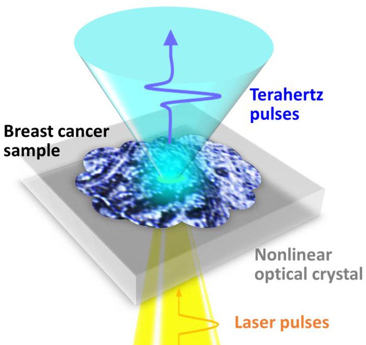
A schematic drawing of the measurement of breast cancer tissue fabricated on a nonlinear optical crystal.
Credit: Osaka University
No stain? No sweat: Terahertz waves can image early-stage breast cancer without staining.
A team of researchers at Osaka University, in collaboration with the University of Bordeaux and the Bergonié Institute in France, has succeeded in terahertz imaging of early-stage breast cancer less than 0.5 mm without staining, which is difficult to identify even by pathological diagnosis. Their work provides a breakthrough towards rapid and precise on-site diagnosis of various types of cancer and accelerates the development of innovative terahertz diagnostic devices.
Breast cancer is roughly divided into two types: invasive and non-invasive. The former, invasive ductal carcinoma (IDC), begins in the cells of a breast duct, growing through the duct walls and into the surrounding breast tissue, potentially spreading to other parts of the body. The latter, ductal carcinoma in situ (DCIS), is an early-stage small breast cancer confined to the breast duct, but it can lead to invasive cancer. Therefore, early detection of DCIS is crucial.
For pathological diagnosis of cancer, the tissue sample is chemically stained, and a pathologist makes a diagnosis using an image of the stained tissue. However, the staining process takes time, and it is difficult to distinguish DCIS from malignant IDC as they look nearly identical.
Terahertz imaging can distinguish cancer tissue from normal tissue without staining and radiation exposure. However, it was still difficult to identify an individual DCIS lesion (which typically range from 50 to 500 μm) by terahertz imaging due to its diffraction-limited spatial resolution of just several millimeters.
“To overcome this drawback, we developed a unique imaging technique in which terahertz light sources that are locally generated at irradiation spots of laser beams in a nonlinear optical crystal directly interact with a breast cancer tissue sample. Consequently, we succeeded in clearly visualizing a DCIS lesion of less than 0.5 mm,” explains lead author Kosuke Okada. The accuracy of this technique is approximately 1000 times higher than that of conventional techniques using terahertz waves.
The researchers also found that terahertz intensity distributions were different between DCIS and IDC, suggesting the possibility of quantitative determination of cancer malignancy.
The breast cancer tissue sample was provided and histologically assessed by collaborators from the University of Bordeaux and the Bergonié Institute. “One of the challenges in this research is preparing a high-quality breast cancer tissue sample fabricated on a nonlinear optical crystal. It is one of the great achievements of international joint research,” says corresponding author Masayoshi Tonouchi.
“Combining our technique with machine learning will aid in the early detection of cancer and determination of cancer malignancy, as well as development of innovative terahertz diagnostic devices using Micro Electro Mechanical Systems.”
###
The article, “Terahertz near-field microscopy of ductal carcinoma in situ (DCIS) of the breast,” will be published online on Oct. 22, 2020 in Journal of Physics: Photonics at DOI:https:/
About Osaka University
Osaka University was founded in 1931 as one of the seven imperial universities of Japan and is now one of Japan’s leading comprehensive universities with a broad disciplinary spectrum. This strength is coupled with a singular drive for innovation that extends throughout the scientific process, from fundamental research to the creation of applied technology with positive economic impacts. Its commitment to innovation has been recognized in Japan and around the world, being named Japan’s most innovative university in 2015 (Reuters 2015 Top 100) and one of the most innovative institutions in the world in 2017 (Innovative Universities and the Nature Index Innovation 2017). Now, Osaka University is leveraging its role as a Designated National University Corporation selected by the Ministry of Education, Culture, Sports, Science and Technology to contribute to innovation for human welfare, sustainable development of society, and social transformation.
Website: https:/
All latest news from the category: Medical Engineering
The development of medical equipment, products and technical procedures is characterized by high research and development costs in a variety of fields related to the study of human medicine.
innovations-report provides informative and stimulating reports and articles on topics ranging from imaging processes, cell and tissue techniques, optical techniques, implants, orthopedic aids, clinical and medical office equipment, dialysis systems and x-ray/radiation monitoring devices to endoscopy, ultrasound, surgical techniques, and dental materials.
Newest articles

Hyperspectral imaging lidar system achieves remote plastic identification
New technology could remotely identify various types of plastics, offering a valuable tool for future monitoring and analysis of oceanic plastic pollution. Researchers have developed a new hyperspectral Raman imaging…

SwRI awarded $26 million to develop NOAA magnetometers
SW-MAG data will help NOAA predict, mitigate the effects of space weather. NASA and the National Oceanic and Atmospheric Administration (NOAA) recently awarded Southwest Research Institute a $26 million contract…

Protein that helps cancer cells dodge CAR T cell therapy
Discovery could lead to new treatments for blood cancer patients currently facing limited options. Scientists at City of Hope®, one of the largest and most advanced cancer research and treatment…


