Navigation software supports kidney research

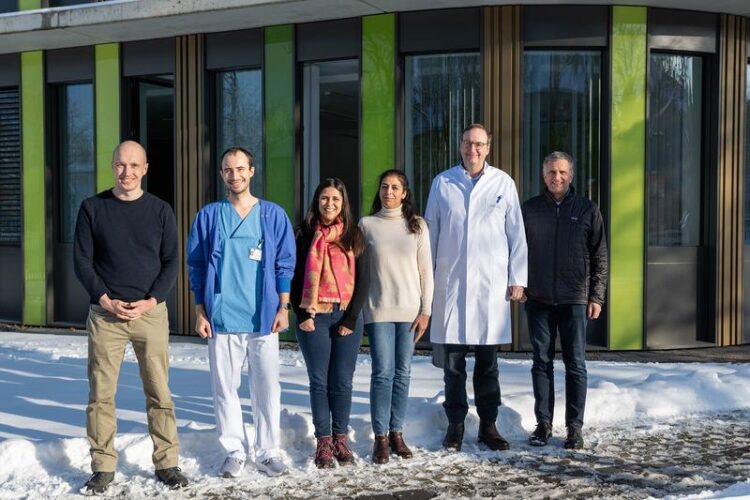
Bonn researchers gain completely new insights into kidney diseases thanks to automated image analysis: (from left): Prof. Alexander Effland, Dr. Alexander Böhner, Alice Jacob, Prof. Zeinab Abdullah, Prof. Christian Kurts and Prof. Martin Rumpf
(c) Alessandro Winkler / University Hospital Bonn (UKB)
Many kidney diseases are manifested by protein in the urine. However, until now it was not possible to determine whether the protein excretion is caused by only a few, but severely damaged, or by many moderately damaged of the millions of small kidney filters, known as glomeruli. Researchers at the University Hospital Bonn, in cooperation with mathematicians from the University of Bonn, have developed a new computer method to clarify this question experimentally. The results of their work have now been published as an article in press in the leading kidney research journal “Kidney International”.
Chronic inflammatory kidney diseases cause serious illnesses, including complete kidney failure, which must be treated with extensive regular blood washing or kidney transplantation. Most of these diseases manifest themselves through protein excretion in the urine. This is because the millions of small filters in the kidneys, known as glomeruli, are damaged and therefore no longer retain protein. It has not yet been possible to determine whether only a few, but severely damaged, or many moderately damaged glomeruli are responsible for protein excretion in the urine. “This is of great importance for therapy and prognosis and could well differ in the many different forms of kidney disease,” says first author Dr. Alexander Böhner, assistant physician at the Clinic for Diagnostic and Interventional Radiology at the UKB, describing the motivation to get to the bottom of this question experimentally in small animal models of kidney disease.
Using the visibility of fluorescent protein to solve puzzles
Behind each glomerulus is a tiny channel in which the urine is processed before it reaches the bladder. If protein is detected in this channel, the associated filter must be defective. The challenge now was to identify this glomerulus. “This was previously an impossible task, because the human kidney consists of millions of glomeruli with tubules that wrap around each other in a complex three-dimensional manner,” says senior author Prof. Christian Kurts from the Institute of Molecular Medicine and Experimental Immunology at the UKB. He is also a member of the Transdisciplinary Research Area 3 (TRA 3) “Life & Health” and the Cluster of Excellence Immunosensation2 at the University of Bonn. The research team solved this problem by making the kidney chemically transparent and imaging it completely using light sheet fluorescence microscopy. The Bonn researchers then combined an image enhancement algorithm with a highly parallelized geometric algorithm that uses a principle similar to that used in navigation devices to determine the fastest route on a two-dimensional map. This made it possible to determine in three dimensions from which defective glomerulus misfiltrated protein originated in a complete kidney. “The algorithm added up all the protein detected in the glomerulus in question and thus determined how much protein it had incorrectly filtered, i.e. how badly damaged it was,” says co-first author Prof. Alexander Effland from the Interdisciplinary Research Unit (IRU) “Mathematics and life sciences” of the Hausdorff Center for Mathematics (HCM) at the University of Bonn, who is also the spokesperson for TRA 1 “Modeling” at the University of Bonn. “The result was a map that shows each glomerulus and its damage in a different color.”
Map of defective kidney filters enables detailed analysis of inflamed kidneys
The Bonn researchers applied this technique to a model of rapid-progressive glomerulonephritis, a particularly aggressive form of kidney inflammation. They found that there are regions in which severely damaged glomeruli are concentrated. This is important for diagnosis by kidney biopsy, in which a sample is taken from the kidney and examined. “If this sample is taken by chance from a severely affected area, the microscopic examination would overestimate the severity of the inflammation of the entire kidney,” summarizes Prof. Kurts. This technique will provide completely new insights into the development of kidney diseases. In principle, it can also be applied to other organs with a modular structure, i.e. consisting of many functional units, such as the liver or lungs.
Promotion
This work was funded by the DFG, in particular the Cluster of Excellence ImmunoSensation2 and the Hausdorff Center for Mathematics at the University of Bonn as well as the Bonfor program for young researchers at the Medical Faculty of Bonn.
Publication: Alexander M. C. Böhner et al; Determining individual glomerular proteinuria and periglomerular infiltration in a cleared murine kidney by 3D fast-marching algorithm, Kidney International; DOI: https://doi.org/10.1016/j.kint.2024.01.043
Press contact:
Dr. Inka Väth
Deputy Press Officer at the University Hospital Bonn (UKB)
Communications and Media Office at Bonn University Hospital
Phone: (+49) 228 287-10596
E-mail: inka.vaeth@ukbonn.de
About Bonn University Hospital: The UKB treats around 500,000 patients per year, employs around 9,000 staff and has total assets of 1.6 billion euros. In addition to the 3,500 medical and dental students, 550 people are trained in numerous healthcare professions each year. The UKB is ranked first among university hospitals (UK) in NRW in the science ranking and in the Focus clinic list and has the third highest case mix index (case severity) in Germany. In 2022 and 2023, the F.A.Z. Institute recognized the UKB as the most desirable employer and training champion among public hospitals in Germany.
Wissenschaftliche Ansprechpartner:
Prof. Dr. Christian Kurts
Institute of Molecular Medicine and Experimental Immunology (IMMEI)
Bonn University Hospital
Phone: +49-(0) 228-287 11050
E-Mail: ckurts@uni-bonn.de
Prof. Alexander Effland
Hausdorff Center for Mathematics (HCM)
University of Bonn
Phone: +49-(0) 228-73 3420
E-mail: effland@iam.uni-bonn.de
Originalpublikation:
Alexander M. C. Böhner et al; Determining individual glomerular proteinuria and periglomerular infiltration in a cleared murine kidney by 3D fast-marching algorithm, Kidney International; DOI: https://doi.org/10.1016/j.kint.2024.01.043
Media Contact
All latest news from the category: Medical Engineering
The development of medical equipment, products and technical procedures is characterized by high research and development costs in a variety of fields related to the study of human medicine.
innovations-report provides informative and stimulating reports and articles on topics ranging from imaging processes, cell and tissue techniques, optical techniques, implants, orthopedic aids, clinical and medical office equipment, dialysis systems and x-ray/radiation monitoring devices to endoscopy, ultrasound, surgical techniques, and dental materials.
Newest articles

First-of-its-kind study uses remote sensing to monitor plastic debris in rivers and lakes
Remote sensing creates a cost-effective solution to monitoring plastic pollution. A first-of-its-kind study from researchers at the University of Minnesota Twin Cities shows how remote sensing can help monitor and…

Laser-based artificial neuron mimics nerve cell functions at lightning speed
With a processing speed a billion times faster than nature, chip-based laser neuron could help advance AI tasks such as pattern recognition and sequence prediction. Researchers have developed a laser-based…

Optimising the processing of plastic waste
Just one look in the yellow bin reveals a colourful jumble of different types of plastic. However, the purer and more uniform plastic waste is, the easier it is to…


