'Neuron-reading' nanowires could accelerate development of drugs for neurological diseases

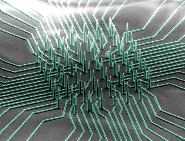
This is a colorized SEM image of the nanowire array. Credit: Integrated Electronics and Biointerfaces Laboratory, UC San Diego
“We're developing tools that will allow us to dig deeper into the science of how the brain works,” said Shadi Dayeh, an electrical engineering professor at the UC San Diego Jacobs School of Engineering and the team's lead investigator.
“We envision that this nanowire technology could be used on stem-cell-derived brain models to identify the most effective drugs for neurological diseases,” said Anne Bang, director of cell biology at the Conrad Prebys Center for Chemical Genomics at the Sanford Burnham Medical Research Institute.
The project was a collaborative effort between the Dayeh and Bang labs, neurobiologists at UC San Diego, and researchers at Nanyang Technological University in Singapore and Sandia National Laboratories. The researchers published their work Apr. 10 in Nano Letters.
Researchers can uncover details about a neuron's health, activity and response to drugs by measuring ion channel currents and changes in its intracellular potential, which is due to the difference in ion concentration between the inside and outside of the cell. The state-of-the-art measurement technique is sensitive to small potential changes and provides readings with high signal-to-noise ratios. However, this method is destructive — it can break the cell membrane and eventually kill the cell. It is also limited to analyzing only one cell at a time, making it impractical for studying large networks of neurons, which are how they are naturally arranged in the body.
“Existing high sensitivity measurement techniques are not scalable to 2D and 3D tissue-like structures cultured in vitro,” Dayeh said. “The development of a nanoscale technology that can measure rapid and minute potential changes in neuronal cellular networks could accelerate drug development for diseases of the central and peripheral nervous systems.”
The nanowire technology developed in Dayeh's laboratory is nondestructive and can simultaneously measure potential changes in multiple neurons — with the high sensitivity and resolution achieved by the current state of the art.
The device consists of an array of silicon nanowires densely packed on a small chip patterned with nickel electrode leads that are coated with silica. The nanowires poke inside cells without damaging them and are sensitive enough to measure small potential changes that are a fraction of or a few millivolts in magnitude. Researchers used the nanowires to record the electrical activity of neurons that were isolated from mice and derived from human induced pluripotent stem cells. These neurons survived and continued functioning for at least six weeks while interfaced with the nanowire array in vitro.
Another innovative feature of this technology is it can isolate the electrical signal measured by each individual nanowire. “This is unusual in existing nanowire technologies, where several wires are electrically shorted together and you cannot differentiate the signal from every single wire,” Dayeh said.
To overcome this hurdle, researchers invented a new wafer bonding approach to fuse the silicon nanowires to the nickel electrodes. Their approach involved a process called silicidation, which is a reaction that binds two solids (silicon and another metal) together without melting either material. This process prevents the nickel electrodes from liquidizing, spreading out and shorting adjacent electrode leads.
Silicidation is usually used to make contacts to transistors, but this is the first time it is being used to do patterned wafer bonding, Dayeh said. “And since this process is used in semiconductor device fabrication, we can integrate versions of these nanowires with CMOS electronics.” Dayeh's laboratory holds several pending patent applications for this technology.
Dayeh noted that the technology needs further optimization for brain-on-chip drug screening. His team is working to extend the application of the technology to heart-on-chip drug screening for cardiac diseases and in vivo brain mapping, which is still several years away due to significant technological and biological challenges that the researchers need to overcome. “Our ultimate goal is to translate this technology to a device that can be implanted in the brain.”
###
A patent is pending for this technology. Contact Skip Cynar in the campus Innovation and Commercialization Office at scynar@ucsd.edu or (858) 822-2672 for licensing information.
Full paper: “High Density Individually Addressable Nanowire Arrays Record Activity from Primary Rodent and Human Stem Cell Derived Neurons.” Authors of the study are Ren Liu, Renjie Chen, Ahmed T. E. Youssef, Sang Heon Lee, Massoud L. Khraiche, John Scott, Yoontae Hwang, Atsunori Tanaka, Yun Goo Ro, Albert K. Matsushita, Xing Dai, Yimin Zhou and Shadi A. Dayeh of UC San Diego; Sandy Hinckley, Deborah Pre, Steven Biesmans and Anne G. Bang of Sanford Burnham Prebys Medical Discovery Institute; Cesare Soci of Nanyang Technological University; and Anthony James, John Nogan, Katherine L. Jungjohann, Douglas V. Pete and Denise B. Webb of Sandia National Laboratories.
This work was supported by a National Science Foundation CAREER award (ECCS-1351980). The team also acknowledges support from the Center for Brain Activity Mapping at UC San Diego, a Calit2 Strategic Research Opportunities award (CITD137) from the Qualcomm Institute at UC San Diego, a Laboratory Directed Research and Development Exploratory Research Award (LDRD-ER) from Los Alamos National Laboratory, the National Institutes of Health (R21 MH099082), a March of Dimes award and a UC San Diego Frontiers of Innovation Scholar Program award. This work was performed in part at UC San Diego's Nanotechnology Infrastructure, a member of the National Nanotechnology Coordinated Infrastructure, which is supported by the National Science Foundation (grant ECCS-1542148).
Media Contact
All latest news from the category: Medical Engineering
The development of medical equipment, products and technical procedures is characterized by high research and development costs in a variety of fields related to the study of human medicine.
innovations-report provides informative and stimulating reports and articles on topics ranging from imaging processes, cell and tissue techniques, optical techniques, implants, orthopedic aids, clinical and medical office equipment, dialysis systems and x-ray/radiation monitoring devices to endoscopy, ultrasound, surgical techniques, and dental materials.
Newest articles

An Endless Loop: How Some Bacteria Evolve Along With the Seasons
The longest natural metagenome time series ever collected, with microbes, reveals a startling evolutionary pattern on repeat. A Microbial “Groundhog Year” in Lake Mendota Like Bill Murray in the movie…

Witness Groundbreaking Research on Achilles Tendon Recovery
Achilles tendon injuries are common but challenging to monitor during recovery due to the limitations of current imaging techniques. Researchers, led by Associate Professor Zeng Nan from the International Graduate…

Why Prevention Is Better Than Cure—A Novel Approach to Infectious Disease Outbreaks
Researchers have come up with a new way to identify more infectious variants of viruses or bacteria that start spreading in humans – including those causing flu, COVID, whooping cough…


