New imaging tracer allows early assessment of abdominal aortic aneurysm risk

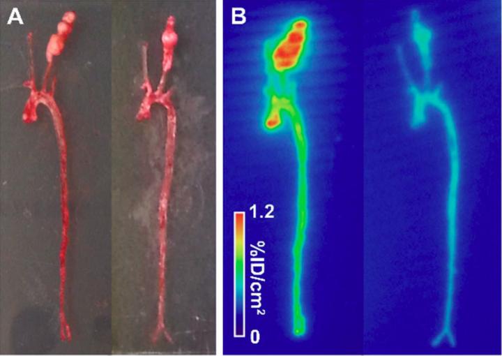
A, B) Examples of ex vivo photography (A) and autoradiography (B) of aortae and carotid arteries from apoE-/-mice with CaCl2-induced carotid aneurysm injected with 99mTc-RYM1 without (left) and with the pre-injection of an excess of MMP inhibitor, RYM (right). Credit: Jakub Toczek, Yunpeng Ye, et al., Yale Cardiovascular Research Center, New Haven, Connecticut
Yale University researchers have developed a way in which medical imaging could potentially be used to assess a patient's rupture risk for abdominal aortic aneurysm. Delaying surgical treatment can be life-threatening, and this new type of imaging could allow physicians to diagnose disease and better plan its management. The study is presented in the featured article of the August issue of The Journal of Nuclear Medicine.
Abdominal aortic aneurysm (AAA) accounts for 10,000 to 15,000 deaths each year in the United States. Matrix metalloproteinases (MMPs) play a key role in the development of AAA, which is especially prevalent in older men with a history of smoking. The researchers developed a novel, water-soluble MMP inhibitor that formed the basis for the new tracer, RYM1. In preclinical evaluation, RYM1–labeled with Tc-99m (99mTc) — was then compared with another MMP tracer in mouse models of aneurysm.
“Studies in mouse models of aneurysm showed that that this tracer allows for imaging vessel wall biology with high sensitivity and specificity, and aortic tracer uptake in vivo correlates with vessel wall inflammation,” explains Mehran M. Sadeghi, MD, of the Yale Cardiovascular Research Center in New Haven and the West Haven VA Medical Center in West Haven, Connecticut.
He points out, “There is no effective medical therapy for AAA, and current guidelines recommend invasive repair of large AAA. However, the morbidity and mortality remain high, so better tools for AAA risk stratification are needed.” The study results show that SPECT/CT imaging with this new tracer, 99mTc-RYM1, is a promising tool for this purpose and could provide new ways to manage AAA.
Looking ahead, Sadeghi says, “Fulfilling the potential of molecular imaging in improving patient care and advancing research is critically dependent on the development of novel tracers with real potential for clinical translation. Remodeling and inflammation are fundamental biological processes which are implicated in the pathogenesis of several diseases, spanning from cancer to heart attack and stroke. MMPs play a key role in tissue remodeling and inflammation. Further development of RYM1-based imaging could expand the applications of molecular imaging and nuclear medicine, and improve patient management in a wide range of diseases.”
###
The authors of “Preclinical evaluation of RYM1, a novel MMP-targeted tracer for imaging aneurysm” include Jakub Toczek, PhD, Yunpeng Ye, PhD, Kiran Gona, PhD, Hye-Yeong Kim, PhD, Jinah Han, PhD, Mahmoud Razavian, PhD, Reza Golestani, MD, PhD, Jiasheng Zhang, MD, Jae-Joon Jung, PhD, and Mehran M. Sadeghi, MD, Cardiovascular Molecular Imaging Laboratory (section of Cardiovascular Medicine) and Yale Cardiovascular Research Center, Yale University School of Medicine, New Haven, Connecticut, and the Veterans Affairs Connecticut Healthcare System, West Haven, Connecticut; and Terence L. Wu, PhD, Yale West Campus Analytical Core, Yale University, West Haven, Connecticut.
This study was supported by grants from the National Institutes of Health (R01-HL112992, R01-HL114703), Connecticut Department of Public Health (2016- 0087) and Department of Veterans Affairs (I0-BX001750).
Please visit the SNMMI Media Center to view the PDF of the study, including images, and more information about molecular imaging and personalized medicine. To schedule an interview with the researchers, please contact Laurie Callahan at (703) 652-6773 or lcallahan@snmmi.org. Current and past issues of The Journal of Nuclear Medicine can be found online at http://jnm.
About the Society of Nuclear Medicine and Molecular Imaging
The Society of Nuclear Medicine and Molecular Imaging (SNMMI) is an international scientific and medical organization dedicated to raising public awareness about nuclear medicine and molecular imaging, a vital element of today's medical practice that adds an additional dimension to diagnosis, changing the way common and devastating diseases are understood and treated and helping provide patients with the best health care possible.
SNMMI's more than 17,000 members set the standard for molecular imaging and nuclear medicine practice by creating guidelines, sharing information through journals and meetings and leading advocacy on key issues that affect molecular imaging and therapy research and practice. For more information, visit http://www.
Media Contact
All latest news from the category: Medical Engineering
The development of medical equipment, products and technical procedures is characterized by high research and development costs in a variety of fields related to the study of human medicine.
innovations-report provides informative and stimulating reports and articles on topics ranging from imaging processes, cell and tissue techniques, optical techniques, implants, orthopedic aids, clinical and medical office equipment, dialysis systems and x-ray/radiation monitoring devices to endoscopy, ultrasound, surgical techniques, and dental materials.
Newest articles
Humans vs Machines—Who’s Better at Recognizing Speech?
Are humans or machines better at recognizing speech? A new study shows that in noisy conditions, current automatic speech recognition (ASR) systems achieve remarkable accuracy and sometimes even surpass human…

Not Lost in Translation: AI Increases Sign Language Recognition Accuracy
Additional data can help differentiate subtle gestures, hand positions, facial expressions The Complexity of Sign Languages Sign languages have been developed by nations around the world to fit the local…

Breaking the Ice: Glacier Melting Alters Arctic Fjord Ecosystems
The regions of the Arctic are particularly vulnerable to climate change. However, there is a lack of comprehensive scientific information about the environmental changes there. Researchers from the Helmholtz Center…


