New PET tracer detects inflammatory arthritis before symptoms appear

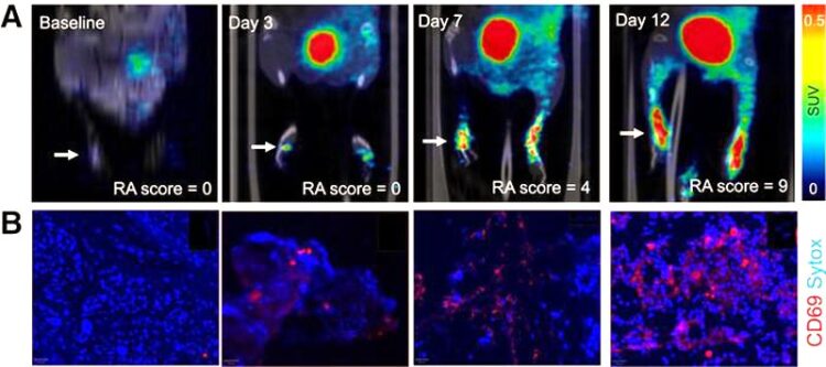
(A) PET images of [68Ga]Ga-DOTA-ZCAM241 uptake at baseline and 3, 7, and 12 days after injection as inflammatory arthritis developed in single representative individual mouse. Images are normalized to SUV of 0.5 for direct comparison between time points. (B) CD69 immunofluorescence Sytox (Thermo Fisher Scientific) staining of joints of representative animals during matching time points.
Image created by E Puuvuori and Y Shen, et al, Uppsala University, Uppsala, Sweden and Karolinska Institutet, Solna, Sweden
A novel PET imaging technique can noninvasively detect active inflammation in the body before clinical symptoms arise, according to research published in the February issue of The Journal of Nuclear Medicine. Using a PET tracer that binds to proteins present on activated immune cells, the technique produces images of ongoing inflammation throughout the body, such as rheumatoid arthritis. This makes it easier for physicians to correctly diagnose and treat patients.
Rheumatoid arthritis is the most common type of inflammatory arthritis and affects 18 million people worldwide. It is a complex autoimmune disease characterized by chronic inflammation. This inflammation can cause the destruction of cartilage and bone, eventually leading to limitations, disabilities, loss of function, decreased quality of life, and possibly shortened life expectancy.
“A major interest of the rheumatology field is employing precision diagnostics to predict disease development in individuals with risk factors of rheumatoid arthritis,” said Fredrik Wermeling, PhD, associate professor and group leader at the Department of Medicine, Division of Rheumatology, Center for Molecular Medicine (CMM) at the Karolinska Institutet, in Solna, Sweden. “The hope is to find ways to identify such individuals even before they get sick, with the goal of being able to treat them so they never develop the disease.”
CD69 is one of the earliest cell surface markers seen on cells experiencing inflammation and is present in the tissue of patients with active rheumatoid arthritis. As such, researchers evaluated the performance of the CD69-targeting PET agent, 68Ga-DOTA-ZCAM241, for early disease detection in a mouse model of inflammatory arthritis.
In the study, mice were imaged with 68Ga-DOTA-ZCAM241 PET before and three, seven, and 12 days after induction of arthritis. Disease progression was monitored by clinical parameters, such as measuring body weight and scoring swelling in the paws. The uptake of 68Ga-DOTA-ZCAM241 in the paws was analyzed, and after the last PET scan, tissue biopsy samples analyzed for CD69 expression. A second group of mice received PET scans with a nonspecific control peptide.
Increased uptake of the CD69-directed tracer 68Ga-DOTA-ZCAM241 was seen in the paws of mice with induced inflammatory arthritis three days after induction, which preceded the appearance of clinical symptoms five to seven days after induction. The uptake of 68Ga-DOTA-ZCAM241 also correlated with the clinical score and disease severity. The nonspecific control peptide demonstrated only low binding.
“68Ga-DOTA-ZCAM241 is a potential candidate for PET imaging of activated immune cells during rheumatoid arthritis onset,” stated Olof Eriksson, PhD, associate professor and group leader of Translational PET Imaging at the Department of Medicinal Chemistry at Uppsala University, in Uppsala, Sweden. “We know that physicians are asking for better methods to image inflammation, for example in rheumatoid arthritis, and we hope this technology will be broadly used in many diseases that involve activated immune cells and inflammation.
This study was made available online in November 2023.
The authors of “Noninvasive PET Detection of CD69-Positive Immune Cells Before Signs of Clinical Disease in Inflammatory Arthritis” include Emma Puuvuori, Gry Hulsart-Billström, Bo Zhang, Pierre Cheung, and Olivia Wegrzyniak, Science for Life Laboratory, Department of Medicinal Chemistry, Uppsala University, Uppsala, Sweden; Yunbing Shen and Fredrik Wermeling, Center for Molecular Medicine, Division of Rheumatology, Department of Medicine, Solna, Karolinska Institutet and Karolinska University Hospital, Stockholm, Sweden; Bogdan Mitran and Olof Eriksson, Department of Medicinal Chemistry, Uppsala University, Uppsala, Sweden, and Antaros Medical AB, Mölndal, Sweden; Sofie Ingvast and Olle Korsgren, Department of Immunology, Genetics and Pathology, Uppsala University, Uppsala, Sweden; Jonas Persson, Science for Life Laboratory, Department of Medicinal Chemistry, Uppsala University, Uppsala, Sweden, and Department of Protein Science, Division of Protein Engineering, KTH Royal Institute of Technology, Stockholm, Sweden; and Stefan Ståhl and John Löfblom, Department of Protein Science, Division of Protein Engineering, KTH Royal Institute of Technology, Stockholm, Sweden.
Visit the JNM website for the latest research, and follow our new Twitter and Facebook pages @JournalofNucMed or follow us on LinkedIn.
Please visit the SNMMI Media Center for more information about molecular imaging and precision imaging. To schedule an interview with the researchers, please contact Rebecca Maxey at (703) 652-6772 or rmaxey@snmmi.org.
About JNM and the Society of Nuclear Medicine and Molecular Imaging
The Journal of Nuclear Medicine (JNM) is the world’s leading nuclear medicine, molecular imaging and theranostics journal, accessed more than 16 million times each year by practitioners around the globe, providing them with the information they need to advance this rapidly expanding field. Current and past issues of The Journal of Nuclear Medicine can be found online at http://jnm.snmjournals.org.
JNM is published by the Society of Nuclear Medicine and Molecular Imaging (SNMMI), an international scientific and medical organization dedicated to advancing nuclear medicine and molecular imaging—precision medicine that allows diagnosis and treatment to be tailored to individual patients in order to achieve the best possible outcomes. For more information, visit www.snmmi.org.
Journal: Journal of Nuclear Medicine
DOI: 10.2967/jnumed.123.266336
Article Title: Noninvasive PET Detection of CD69-Positive Immune Cells Before Signs of Clinical Disease in Inflammatory Arthritis
Article Publication Date: 1-Feb-2024
COI Statement: John Löfblom, Jonas Persson, Stefan Ståhl, Olof Eriksson, and Olle Korsgren are inventors of a patent covering ZCAM241. Bogdan Mitran and Olof Eriksson are employees of Antaros Medical AB. Olof Eriksson and Olle Korsgren are shareholders of Antaros Tracer AB.
Media Contact
Rebecca Maxey
Society of Nuclear Medicine and Molecular Imaging
rmaxey@snmmi.org
Office: 703-652-6772
All latest news from the category: Medical Engineering
The development of medical equipment, products and technical procedures is characterized by high research and development costs in a variety of fields related to the study of human medicine.
innovations-report provides informative and stimulating reports and articles on topics ranging from imaging processes, cell and tissue techniques, optical techniques, implants, orthopedic aids, clinical and medical office equipment, dialysis systems and x-ray/radiation monitoring devices to endoscopy, ultrasound, surgical techniques, and dental materials.
Newest articles

An Endless Loop: How Some Bacteria Evolve Along With the Seasons
The longest natural metagenome time series ever collected, with microbes, reveals a startling evolutionary pattern on repeat. A Microbial “Groundhog Year” in Lake Mendota Like Bill Murray in the movie…

Witness Groundbreaking Research on Achilles Tendon Recovery
Achilles tendon injuries are common but challenging to monitor during recovery due to the limitations of current imaging techniques. Researchers, led by Associate Professor Zeng Nan from the International Graduate…

Why Prevention Is Better Than Cure—A Novel Approach to Infectious Disease Outbreaks
Researchers have come up with a new way to identify more infectious variants of viruses or bacteria that start spreading in humans – including those causing flu, COVID, whooping cough…


