Novel PET tracer identifies most bacterial infections

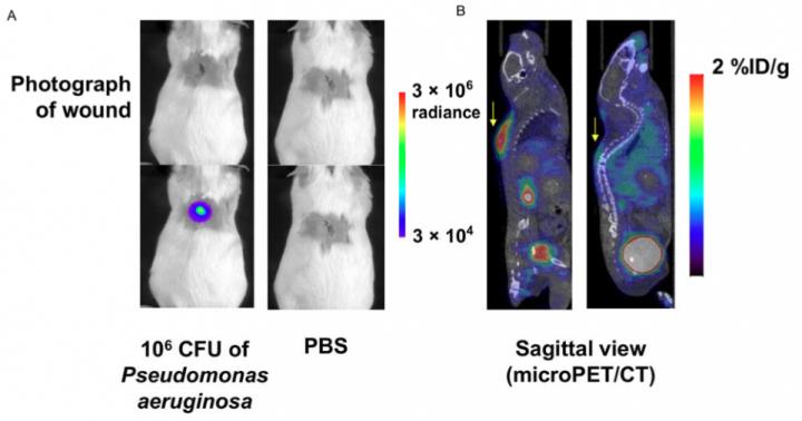
This figure shows A) bioluminescence images of CD1 mice bearing P. aeruginosa-infected wound (left panel) and control mice (right panel), and B) sagittal slices from micro PET/CT scan of the same mice 1 hour after intravenous administration of 6"-<sup>18</sup>F-fluoromaltotriose. Courtesy of Sam Gambhir, MD, PhD, Stanford University
Stanford University medical scientists have developed a novel imaging agent that could be used to identify most bacterial infections. The study is the featured basic science article in The Journal of Nuclear Medicine's October issue.
Bacteria are good at mutating to become resistant to antibiotics. As one way to combat the problem of antimicrobial resistance, the Centers for Disease Control and Prevention (CDC) has called for the development of novel diagnostics to detect and help manage the treatment of infectious diseases.
“We really lack tools in the clinic to be able to visualize bacterial infections,” explains Sanjiv Sam Gambhir, MD, PhD, chair of the Radiology Department and director of Precision Health and Integrated Diagnostics at Stanford University in California.
“What we need is something that bacteria eat that your cells, so-called mammalian cells, do not. As it turns out, there is such an agent, and that agent is maltose, which is taken up only by bacteria because they have a transporter, called a maltodextrine transporter, on their cell wall that is able to take up maltose and small derivatives of maltose.”
The traditional way of diagnosing bacterial infection involves biopsy of the infected tissue and/or blood and culture tests. Gambhir and colleagues developed a new positron emission tomography (PET) tracer, 6″-18F-fluoromaltotriose, that offers a non-invasive means of detection.
The agent is a derivative of maltose and is labeled with radioactive fluorine-18 (18F). For this study, the tracer was evaluated in several clinically relevant bacterial strains in cultures and in mouse models using a micro-PET/CT scanner. Its use to help monitor antibiotic therapies was also evaluated in rats.
The results show that 6″-18F-fluoromaltotriose was taken up in both gram-positive and gram-negative bacterial strains, and it was able to detect Pseudomonas aeruginosa in a clinically relevant mouse model of wound infection.
Gambhir points out, “This is the first time this particular maltotriose, labeled with fluorine-18, has been synthesized and used in animal models. It's able to pick up bacteria that may be present anywhere throughout your body, and it does not lead to an imaging signal from a site of infection that does not involve bacteria.”
He notes that the new agent even identified an infection in the heart of an animal. “We could pick up very small bacterial foci in a heart valve. And then when those animals were treated with an antibiotic, we could see that the signal went away in the heart. So, the properties of the tracer of sensitivity, specificity, low background signal throughout the animal are now facilitating its translation into humans.”
The results of this pre-clinical study demonstrate that 6″-18F-fluoromaltotriose is a promising new tracer for diagnosing most bacterial infections and has the potential to change the clinical management of patients suffering from infectious diseases of bacterial origin.
Looking ahead, Gambhir says, “The hope is that in the future when someone has a potential infection, this approach of injecting the patient with fluoromaltotriose and imaging them in a PET scanner will allow localization of the signal and, therefore, the bacteria. And then, as one treats them, one can verify that the treatment is actually working – so that if it's not working, one can quickly change to a different treatment (for example, a different antibiotic). These kinds of findings are very important for patients, because they will very likely lead to entirely new ways to manage patients with bacterial infections, no matter where those infections might be hiding in the body.”
###
The authors of “Specific Imaging of Bacterial Infection using 6”-18F-Fluoromaltotriose: A Second-Generation PET Tracer Targeting the Maltodextrin Transporter in Bacteria” include Gayatri Gowrishankar, Jonathan Hardy, Mirwais Wardak, Mohammad Namavari, Robert E. Reeves, Evgenios Neofytou, Ananth Srinivasan, Joseph C. Wu, Christopher H. Contag, and Sanjiv Sam Gambhir, Stanford University School of Medicine, Stanford, California.
Please visit the SNMMI Media Center to view the PDF of the study, including images, and more information about molecular imaging and personalized medicine. To schedule an interview with the researchers, please contact Laurie Callahan at 703-652-6773 or lcallahan@snmmi.org. Current and past issues of The Journal of Nuclear Medicine can be found online at http://jnm.
About the Society of Nuclear Medicine and Molecular Imaging
The Society of Nuclear Medicine and Molecular Imaging (SNMMI) is an international scientific and medical organization dedicated to raising public awareness about nuclear medicine and molecular imaging, a vital element of today's medical practice that adds an additional dimension to diagnosis, changing the way common and devastating diseases are understood and treated and helping provide patients with the best health care possible.
SNMMI's more than 15,000 members set the standard for molecular imaging and nuclear medicine practice by creating guidelines, sharing information through journals and meetings and leading advocacy on key issues that affect molecular imaging and therapy research and practice. For more information, visit http://www.
Media Contact
All latest news from the category: Medical Engineering
The development of medical equipment, products and technical procedures is characterized by high research and development costs in a variety of fields related to the study of human medicine.
innovations-report provides informative and stimulating reports and articles on topics ranging from imaging processes, cell and tissue techniques, optical techniques, implants, orthopedic aids, clinical and medical office equipment, dialysis systems and x-ray/radiation monitoring devices to endoscopy, ultrasound, surgical techniques, and dental materials.
Newest articles
Humans vs Machines—Who’s Better at Recognizing Speech?
Are humans or machines better at recognizing speech? A new study shows that in noisy conditions, current automatic speech recognition (ASR) systems achieve remarkable accuracy and sometimes even surpass human…

Not Lost in Translation: AI Increases Sign Language Recognition Accuracy
Additional data can help differentiate subtle gestures, hand positions, facial expressions The Complexity of Sign Languages Sign languages have been developed by nations around the world to fit the local…

Breaking the Ice: Glacier Melting Alters Arctic Fjord Ecosystems
The regions of the Arctic are particularly vulnerable to climate change. However, there is a lack of comprehensive scientific information about the environmental changes there. Researchers from the Helmholtz Center…


