PET radiotracer design for monitoring targeted immunotherapy

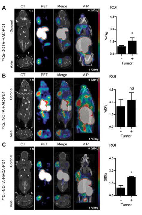
Comparison of (A) Cu-64-DOTA-HACPD1, (B) Cu-64-NOTA-HAC-PD1, and (C) Cu-64-NOTA-HACA-PD1 ImmunoPET images acquired at 1h p.i. (~1.85 MBq/10 μg/200 μl) of NSG mice bearing dual subcutaneous tumors in shoulders (right = hPD-L1+; left = hPD-L1-). Panels left to right show representative coronal and axial cross sections of CT, PET, and merged PET/CT images with maximum intensity projection (MIP). White dashed line, coronal image, demarcation of axial cross-section; White dashed line, axial image, tumor boundary. SG, salivary gland; T+, hPD-L1 positive tumor; T-, hPD-L1 negative tumor; L, lung; Li, liver; K, kidney; B, bladder; H, heart. Scale bars represent 1 percent (blue) - 6 percent (red) ID/g. Tumor uptake was quantified using ROI analysis (right panel) without partial volume correction. Error bars represent SD. ns, not significant; *p<0.05, **p<0.01, ***p<0.001, student's T test. Credit: Sam Gambhir, MD, PhD, Stanford University
A drug that blocks a cancer's inhibitory checkpoint molecules is called an immune checkpoint inhibitor and this form of immunotherapy has emerged as a promising cancer treatment approach. However, the lack of imaging tools to assess immune checkpoint expression has been a major barrier to predicting and monitoring response to a clinical checkpoint blockade.
“Because immunotherapies for cancer are expanding, methods to optimize them for an individual patient through molecular imaging are needed,” explains Sam Gambhir, MD, PhD, at Stanford University. “Using animal models, this study shows the development of several new engineered PET tracers that can help image the immune system in action and be used to monitor checkpoint inhibitor therapy.”
The study assessed practical immunoPET radiotracer design modifications and their effects on human PD-L1 immune checkpoint imaging. The researchers sought to optimize engineering design parameters including chelate, glycosylation, and radiometal to develop a non-invasive molecular imaging tool for eventual monitoring of clinical checkpoint blockade.
Gambhir points out, “This research will ultimately allow for translation to human imaging of the tracer that worked best in animal models. An effective immunoPET tracer will help patients receiving checkpoint inhibitor immunotherapy get optimal treatment and have the best chance for fighting their cancer.”
Molecular imaging is playing an increasingly integral role in immunotherapy and personalized medicine. Looking ahead, Gambhir envisions “a lot more use of PET/CT or PET/MR imaging for patients undergoing immunotherapy.” He adds, “This and related research will also help us develop other imaging approaches for understanding the immune system in action.”
###
The authors of “Practical ImmunoPET radiotracer design considerations for human immune checkpoint imaging” include Aaron T. Mayer, Arutselvan Natarajan, Sydney R. Gordon, Roy L. Maute, Melissa N. McCracken, Aaron M. Ring, Irving L. Weissman and Sanjiv S. Gambhir, Stanford University, Stanford, California.
Support for this study was provided by Kenneth Lau, Frezghi Habte, the Canary Foundation and the Ben and Catherine Ivy Foundation. This material is based upon work supported by a National Science Foundation Graduate Research Fellowship Grant (DGE-114747) and a NIH TBi2 Training Grant (2T32EB009653-06). MicroPET/CT imaging and Gamma Counter measurements were performed in the SCi3 Stanford Small Animal Imaging Service Center.
Please visit the SNMMI Media Center to view the PDF of the study, including images, and more information about molecular imaging and personalized medicine. To schedule an interview with the researchers, please contact Laurie Callahan at (703) 652-6773 or lcallahan@snmmi.org. Current and past issues of The Journal of Nuclear Medicine can be found online at http://jnm.
About the Society of Nuclear Medicine and Molecular Imaging
The Society of Nuclear Medicine and Molecular Imaging (SNMMI) is an international scientific and medical organization dedicated to raising public awareness about nuclear medicine and molecular imaging, a vital element of today's medical practice that adds an additional dimension to diagnosis, changing the way common and devastating diseases are understood and treated and helping provide patients with the best health care possible.
SNMMI's more than 17,000 members set the standard for molecular imaging and nuclear medicine practice by creating guidelines, sharing information through journals and meetings and leading advocacy on key issues that affect molecular imaging and therapy research and practice. For more information, visit http://www.
Media Contact
All latest news from the category: Medical Engineering
The development of medical equipment, products and technical procedures is characterized by high research and development costs in a variety of fields related to the study of human medicine.
innovations-report provides informative and stimulating reports and articles on topics ranging from imaging processes, cell and tissue techniques, optical techniques, implants, orthopedic aids, clinical and medical office equipment, dialysis systems and x-ray/radiation monitoring devices to endoscopy, ultrasound, surgical techniques, and dental materials.
Newest articles

Single-Celled Heroes: Foraminifera’s Power to Combat Ocean Phosphate Pollution
So-called foraminifera are found in all the world’s oceans. Now an international study led by the University of Hamburg has shown that the microorganisms, most of which bear shells, absorb…

Humans vs Machines—Who’s Better at Recognizing Speech?
Are humans or machines better at recognizing speech? A new study shows that in noisy conditions, current automatic speech recognition (ASR) systems achieve remarkable accuracy and sometimes even surpass human…

Not Lost in Translation: AI Increases Sign Language Recognition Accuracy
Additional data can help differentiate subtle gestures, hand positions, facial expressions The Complexity of Sign Languages Sign languages have been developed by nations around the world to fit the local…


