Smart contact lenses for cancer diagnostics and screening

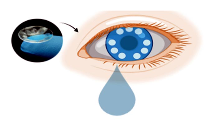
Scientists from the Terasaki Institute for Biomedical Innovation (TIBI) have developed a contact lens that can capture tears for the detection of exosomes, nanometer-sized vesicles found in bodily secretions which have the potential for being diagnostic cancer biomarkers.
Credit: Terasaki Institute for Biomedical Innovation, using Biorender software
Scientists from the Terasaki Institute for Biomedical Innovation (TIBI) have developed a contact lens that can capture and detect exosomes, nanometer-sized vesicles found in bodily secretions which have the potential for being diagnostic cancer biomarkers. The lens was designed with microchambers bound to antibodies that can capture exosomes found in tears. This antibody- conjugated signaling microchamber contact lens (ACSM-CL) can be stained for detection with nanoparticle-tagged specific antibodies for selective visualization. This offers a potential platform for cancer pre-screening and a supportive diagnostic tool that is easy, rapid, sensitive, cost-effective, and non-invasive.
Exosomes are formed within most cells and secreted into many bodily fluids, such as plasma, saliva, urine, and tears. Once thought to be the dumping grounds for unwanted materials from their cells of origin, it is now known that exosomes can transport different biomolecules between cells. It has also been shown that there is a wealth of surface proteins on exosomes – some that are common to all exosomes and others that are increased in response to cancer, viral infections, or injury. In addition, exosomes derived from tumors can strongly influence tumor regulation, progression, and metastasis.
Because of these capabilities, there has been much interest in using exosomes for cancer diagnosis and prognosis/treatment prediction. However, this has been hampered by the difficulty in isolating exosomes in sufficient quantity and purity for this purpose. Current methods involve tedious and time-consuming ultracentrifuge and density gradients, lasting at least ten hours to complete. Further difficulties are posed in detection of the isolated exosomes; commonly used methods require expensive and space-consuming equipment.
The TIBI team has leveraged their expertise in contact lens biosensor design and fabrication to eliminate the need for these isolation methods by devising their ACSM-CL for capturing exosomes from tears, an optimum and cleaner source of exosomes than blood, urine, and saliva.
They also facilitated and optimized the preparation of their ACSM-CL by the use of alternative approaches. When fabricating the microchambers for their lens, the team used a direct laser cutting and engraving approach rather than conventional cast molding for structural retention of both the chambers and the lens.
In addition, the team introduced a method that chemically modified the microchamber surfaces to activate them for antibody binding. This method was used in place of standard approaches, in which metallic or nanocarbon materials must be used in expensive clean-room settings.
The team then optimized procedures for binding a capture antibody to the ACSM-CL microchambers and a different (positive control) detection antibody onto gold nanoparticles that can be visualized spectroscopically. Both these antibodies are specific for two different surface markers found on all exosomes.
In an initial validation experiment, the ACSM-CL was tested against exosomes secreted into supernatants from ten different tissue and cancer cell lines. The ability to capture and detect exosomes was validated by the spectroscopic shifts observed in all the test samples, in comparison with the negative controls. Similar results were obtained when the ACSM-CL was tested against ten different tear samples collected from volunteers.
In final experiments, exosomes in supernatants collected from three different cell lines with different surface marker expressions were tested against the ACSM-CL, along with different combinations of marker-specific detection antibodies. The resultant patterns of detection and non-detection of exosomes from the three different cell lines were as expected, thus validating the ACSM-CL’s ability to accurately capture and detect exosomes with different surface markers.
“Exosomes are a rich source of markers and biomolecules which can be targeted for several biomedical applications,” said Ali Khademhosseini, Ph.D., TIBI’s Director and CEO. “The methodology that our team has developed greatly facilitates our ability to tap into this source.”
Additional authors are: Shaopei Li, Yangzhi Zhu, Reihaneh Haghniaz, Satoru Kawakita, Shenghan Guan, Jianjun Chen, Kalpana Mandal, Juchen Guo, Heemin Kang, Wujin Sun, Han-Jun Kim, Vadim Jucaud, Mehmet R. Dokmeci, Pete Kollbaum, Chi Hwan Lee, and Ali Khademhosseini.
The Terasaki Institute for Biomedical Innovation (terasaki.org) is a non-profit research organization that invents and fosters practical solutions that restore or enhance the health of individuals. Research at the Terasaki Institute leverages scientific advancements that enable an understanding of what makes each person unique, from the macroscale of human tissues down to the microscale of genes, to create technological solutions for some of the most pressing medical problems of our time. We use innovative technology platforms to study human disease on the level of individual patients by incorporating advanced computational and tissue-engineering methods. Findings yielded by these studies are translated by our research teams into tailored diagnostic and therapeutic approaches encompassing personalized materials, cells and implants with unique potential and broad applicability to a variety of diseases, disorders and injuries.
The Institute is made possible through an endowment from the late Dr. Paul I Terasaki, a pioneer in the field of organ transplant technology.
Journal: Advanced Functional Materials
DOI: 10.1002/adfm.202206620
Method of Research: Experimental study
Subject of Research: Not applicable
Article Title: A microchambers containing contact lens for the non-invasive detection of tear exosomes
Article Publication Date: 10-Aug-2022
Media Contact
Stewart Han
Terasaki Institute for Biomedical Innovation
shan@terasaki.org
Office: 818-836-4393
All latest news from the category: Medical Engineering
The development of medical equipment, products and technical procedures is characterized by high research and development costs in a variety of fields related to the study of human medicine.
innovations-report provides informative and stimulating reports and articles on topics ranging from imaging processes, cell and tissue techniques, optical techniques, implants, orthopedic aids, clinical and medical office equipment, dialysis systems and x-ray/radiation monitoring devices to endoscopy, ultrasound, surgical techniques, and dental materials.
Newest articles

First-of-its-kind study uses remote sensing to monitor plastic debris in rivers and lakes
Remote sensing creates a cost-effective solution to monitoring plastic pollution. A first-of-its-kind study from researchers at the University of Minnesota Twin Cities shows how remote sensing can help monitor and…

Laser-based artificial neuron mimics nerve cell functions at lightning speed
With a processing speed a billion times faster than nature, chip-based laser neuron could help advance AI tasks such as pattern recognition and sequence prediction. Researchers have developed a laser-based…

Optimising the processing of plastic waste
Just one look in the yellow bin reveals a colourful jumble of different types of plastic. However, the purer and more uniform plastic waste is, the easier it is to…


