Ultra-low dose total body PET/CT effective for evaluating arthritis

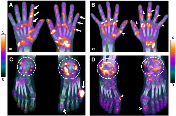
18F-FDG uptake in hands and feet of participants with AIA. (A) A 54-y-old man with PsA showing elevated uptake at multiple hand joints. Raylike distribution, as indicated by arrows, in metacarpophalangeal (MCP), proximal interphalangeal (PIP), and distal interphalangeal (DIP) joints in sequence, attributed to involvement of flexor or extensor tendons. (B) A 47-y-old woman with RA, showing involvement of entire row of MCP (arrowheads, right hand) and PIP/interphalangeal (IP) joints (arrowheads, left hand). (C) Feet images of same PsA participant in A, demonstrating increased uptake at ankle joints (dashed circles), more intense on the left side, and left first IP and fifth metatarsophalangeal (MTP) joints (arrows). (D) Feet images of a 71-y-old man with RA, demonstrating bilateral, rather symmetric, uptake around ankles as well as right first MTP and left first IP joints, suggestive of synovitis (arrowheads).
Image created by Y Abdelhafez and A Chaudhari et al, University of California, Davis, Sacramento, CA, USA
Total body PET/CT scans can successfully visualize systemic joint involvement in patients with autoimmune arthritis, according to new first-in-human research published in the October issue of The Journal of Nuclear Medicine. The total body PET/CT scans showed high agreement with standard joint-by-joint rheumatological evaluation and a moderate to strong correlation with rheumatological outcome measures.
Autoimmune inflammatory arthritides (AIA)—such as psoriatic arthritis and rheumatoid arthritis—are chronic, systemic conditions that cause joint inflammation, joint destruction and pain. According to the Centers for Disease Control and Prevention, approximately one in four adults, or 58 million Americans, have been diagnosed with arthritis by a doctor. By 2040, an estimated 78 million adults are projected to be diagnosed with arthritis.
“Currently, there are significant clinical challenges in managing AIA populations. For example, it is unclear which patients should receive which treatments, how exactly these treatments change the inflammatory status of different tissues or outcomes, and the impact the disease and treatments have on other organs of the body,” said Abhijit J. Chaudhari, PhD, professor of radiology at the University of California–Davis in Davis, California. “Systemic molecular imaging enabled by total body PET could provide currently unavailable, objective biomarkers that could help address these challenges.”
To assess the performance of molecular imaging to evaluate AIA, researchers used an ultra-low dose 18F-FDG total-body PET/CT acquisition protocol to scan 30 participants (24 with AIA and six with osteoarthritis). Participants also underwent a joint-by-joint rheumatological evaluation. In total, 1,997 joints were evaluated.
Total body PET/CT was successful in visualizing 18F-FDG uptake at joints throughout the entire body, including those of the hands and feet, without the need for custom positioning of the participants. In the AIA cohort, there was concordance between total body PET qualitative assessments and joint-by-joint rheumatological evaluation for 69.9 percent of the participants. About 20 percent of the AIA group had joints that were deemed negative on rheumatological evaluation but were positive on PET/CT, and 10 percent were negative on PET/CT but positive on the rheumatological evaluation. For the osteoarthritis cohort, there was agreement in joint assessment among 91.1 percent of participants. In this group 8.8 percent of joints were deemed negative on rheumatological evaluation but were positive on PET/CT, and no joints were negative on PET/CT but positive on rheumatological exam.
“Systemic evaluation of arthritic disease activity across all musculoskeletal tissues of the body may provide unique insights for assessing disease burdenrisk stratification, treatment selection, and monitoring on treatment response. The biomarkers imaged may also have clear potential to accelerate arthritic drug discovery and development,” noted Chaudhari. “In addition, the inflammatory arthritides assessed in the paper are part of a broad category of autoimmune disorders. This work may contribute to improved understanding of total-body impact of autoimmunity.”
The authors of “Total-Body 18F-FDG PET/CT in Autoimmune Inflammatory Arthritis at Ultra-Low Dose: Initial Observations” include Yasser Abdelhafez, Department of Radiology, University of California Davis, Davis, California, and Nuclear Medicine Unit, South Egypt Cancer Institute, Assiut University, Egypt; Siba P. Raychaudhuri, Department of Internal Medicine-Rheumatology, University of California Davis, Davis, California, and Northern California Veterans Affairs Medical Center, Mather, California; Dario Mazza, Heather L. Hunt, Kristin McBride, Mike Nguyen, Denise T. Caudle, Lorenzo Nardo and Abhijit J. Chaudhari, Department of Radiology, University of California Davis, Davis, California; Soumajyoti Sarkar, Department of Internal Medicine-Rheumatology, University of California Davis, Davis, California; Benjamin A. Spencer, Negar Omidvari, Simon Cherry and Ramsey D. Badawi, Department of Radiology, University of California Davis, Davis, California, and Department of Biomedical Engineering, University of California Davis, Davis, California; and Heejung Bang, Department of Public Health Sciences, University of California Davis, Davis, California.
Visit the JNM website for the latest research, and follow our new Twitter and Facebook pages @JournalofNucMed or follow us on LinkedIn.
Please visit the SNMMI Media Center for more information about molecular imaging and precision imaging. To schedule an interview with the researchers, please contact Rebecca Maxey at (703) 652-6772 or rmaxey@snmmi.org.
About JNM and the Society of Nuclear Medicine and Molecular Imaging
The Journal of Nuclear Medicine (JNM) is the world’s leading nuclear medicine, molecular imaging and theranostics journal, accessed 15 million times each year by practitioners around the globe, providing them with the information they need to advance this rapidly expanding field. Current and past issues of The Journal of Nuclear Medicine can be found online at http://jnm.snmjournals.org.
JNM is published by the Society of Nuclear Medicine and Molecular Imaging (SNMMI), an international scientific and medical organization dedicated to advancing nuclear medicine and molecular imaging—precision medicine that allows diagnosis and treatment to be tailored to individual patients in order to achieve the best possible outcomes. For more information, visit www.snmmi.org.
Journal: Journal of Nuclear Medicine
DOI: 10.2967/jnumed.121.263774
Article Title: Total-Body 18F-FDG PET/CT in Autoimmune Inflammatory Arthritis at Ultra-Low Dose: Initial Observations
Article Publication Date: 1-Oct-2022
COI Statement: University of California Davis has a research and a revenue-sharing agreement with United Imaging Health Care. Ramsey D. Badawi, Simon R. Cherry, and Lorenzo Nardo are investigators on a research grant funded by United Imaging Health Care, the manufacturer of the scanner used in this article.
Media Contact
Rebecca Maxey
Society of Nuclear Medicine and Molecular Imaging
rmaxey@snmmi.org
Office: 703-652-6772
All latest news from the category: Medical Engineering
The development of medical equipment, products and technical procedures is characterized by high research and development costs in a variety of fields related to the study of human medicine.
innovations-report provides informative and stimulating reports and articles on topics ranging from imaging processes, cell and tissue techniques, optical techniques, implants, orthopedic aids, clinical and medical office equipment, dialysis systems and x-ray/radiation monitoring devices to endoscopy, ultrasound, surgical techniques, and dental materials.
Newest articles

Innovative 3D printed scaffolds offer new hope for bone healing
Researchers at the Institute for Bioengineering of Catalonia have developed novel 3D printed PLA-CaP scaffolds that promote blood vessel formation, ensuring better healing and regeneration of bone tissue. Bone is…

The surprising role of gut infection in Alzheimer’s disease
ASU- and Banner Alzheimer’s Institute-led study implicates link between a common virus and the disease, which travels from the gut to the brain and may be a target for antiviral…

Molecular gardening: New enzymes discovered for protein modification pruning
How deubiquitinases USP53 and USP54 cleave long polyubiquitin chains and how the former is linked to liver disease in children. Deubiquitinases (DUBs) are enzymes used by cells to trim protein…


