Portable CT Scanner Joins Hunt for Alternative Energy

Berkeley Lab’s portable scanner has sailed the high seas and endured arctic cold, imaging more than 2000 feet of core sample along the way
Lawrence Berkeley National Laboratory (Berkeley Lab) scientists have developed the world’s first x-ray computed tomography (CT) scanner capable of examining entire core samples at remote drilling sites. The portable device, which employs the same high-resolution imaging technology used to diagnose diseases, could help researchers determine how to best extract the vast quantities of natural gas hidden under the world’s oceans and permafrost.
The scanner images the distribution of gas hydrates in core samples pulled from deeply buried sediment. These hydrates are a latticework of water and methane that form an ice-like solid under high pressures and temperatures that hover just above freezing, conditions found in deep oceans and under Arctic permafrost. Scientists estimate the methane trapped in this crystalline mix may yield far more energy than the planet’s remaining reserves of fossil fuel.
But they must first determine how to find and remove it. As part of this investigational legwork, researchers drill into likely gas hydrate reserves and extract core samples. Select samples are then shipped to laboratories for analysis, and the resulting data is used to develop computer models that predict how gas hydrates behave in sediments, which may help researchers determine how to most efficiently locate and extract methane.
It’s a laborious process, however. Because gas hydrates rapidly decompose when brought to the surface, the samples must be preserved under high pressure and low temperatures, then shipped to labs hundreds of miles away. This means the data required for these powerful numerical models is harvested slowly, one carefully packaged-and-shipped core at a time.
Barry Freifeld, a mechanical engineer in Berkeley Lab’s Earth Sciences Division, wondered if real-time, on-site analysis could expedite this work. His optimism stemmed from earlier research in which he demonstrated that a medical CT scanner can image a wave of methane hydrate dissociating in a sand mixture.
“Nobody had ever done that before, and I asked why can’t we also do it in the field,” Freifeld says.
Unfortunately, most CT scanners weigh more than 1000 kilograms, are bolted to the floor, and are housed in lead-lined rooms. Portable they’re not. On the other hand, their ability to splice hundreds of x-ray scans into one cross-sectional image could enable researchers to map the distribution of gas hydrates in core samples in unprecedented detail. If only such power could be reduced in size and brought to the drill site.
Freifeld believed he knew how, and he got his break last spring after learning the drill ship JOIDES Resolution was scheduled to probe for gas hydrates off the Oregon coast. The vessel is operated by the Ocean Drilling Program, an international partnership of scientists and research institutions sponsored by the National Science Foundation and participating countries. The group had previously used conventional x-ray imaging aboard the ship to analyze core samples, but the images proved of marginal quality. When Freifield suggested x-ray CT, essentially offering laboratory-quality analysis on the high seas, the Ocean Drilling Program jumped at the chance.
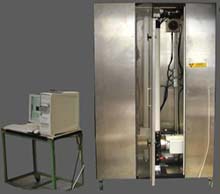
His team received funding from the Department of Energy’s National Energy Technology Laboratory, and in five weeks built a refrigerator-sized, 300-kilogram scanner. They trucked it to Oregon’s Coos Bay, loaded it on a supply ship, sailed west overnight, and at daybreak hoisted it aboard the JOIDES Resolution as it drilled along the Cascadia Ridge in search of hydrates. Several hours later, the scanner analyzed its first core sample, and churned through 1500 feet of core over the next several weeks.
“We can run core through the scanner almost as quickly as they can pull it out,” says Freifeld “Now, researchers don’t have to send kilometers of core to a lab to get the same information they can obtain in the field. They’ll send data instead of rocks.”
Their success hinges on several innovations. Instead of a lead-lined room to protect operators from radiation, they developed a three-piece shield composed of a layer of lead sandwiched between two thin stainless steel layers. This arrangement reduces the amount of lead usually required to encapsulate x-ray imaging systems. And because x-rays passing through the center of the core are more attenuated than those passing through the edges, they designed a half-cylinder-shaped, aluminum compensator that flattens the image intensity and ensures high-resolution imaging throughout the core sample. In addition, special software reconstructs a 3-D image of a scanned core, giving an operator the freedom to observe the core’s interior from any angle and direction. And 3-D scans can be taken at a rate of three minutes per foot of core length, yielding resolutions between 50 and 200 microns.
“We’ve taken a million dollar medical instrument and transformed it into a rugged, $150,000 piece of equipment,” Freifeld says.
This winter, the hearty scanner traveled above the Arctic Circle to the permafrost stretches near Prudhoe Bay, Alaska. There, researchers are conducting the first test on U.S. soil concerning how to extract methane from gas hydrates. The scanner analyzed more than 500 feet of core sample, enabling researchers to generate the most detailed log of permafrost cores ever recorded. And the system worked in subzero temperatures.
“It ran fine, but the cold was hard on the technicians. We needed a lot of tea and coffee,” Freifeld says.
Luckily for Freifeld, the scanner is next headed to warmer climates. It’s scheduled for another hitch aboard the JOIDES Resolution as it sails from Bermuda to Newfoundland. The ship will drill along the continental margin and study rifting, the tectonic process by which the lithosphere thins and the seafloors spread. The scanner will allow scientists to generate the most detailed lithostratigraphic record ever constructed from oceanic cores.
“With this instrument, we can systematically investigate everything recovered and generate a detailed electronic record,” Freifeld says. “Its ability to conduct high-resolution imaging anywhere will have a large impact on energy exploration, mining and fundamental research.”
In addition to Freifeld, Tim Kneafsey, Jacob Pruess, Paul Reiter, and Liviu Tomutsa of Berkeley Lab’s Earth Sciences Division contributed to the development of the scanner.
Berkeley Lab is a U.S. Department of Energy national laboratory located in Berkeley, California. It conducts unclassified scientific research and is managed by the University of California.
Media Contact
More Information:
http://www.lbl.gov/Science-Articles/Archive/ESD-CT-scanner.htmlAll latest news from the category: Power and Electrical Engineering
This topic covers issues related to energy generation, conversion, transportation and consumption and how the industry is addressing the challenge of energy efficiency in general.
innovations-report provides in-depth and informative reports and articles on subjects ranging from wind energy, fuel cell technology, solar energy, geothermal energy, petroleum, gas, nuclear engineering, alternative energy and energy efficiency to fusion, hydrogen and superconductor technologies.
Newest articles

Innovative 3D printed scaffolds offer new hope for bone healing
Researchers at the Institute for Bioengineering of Catalonia have developed novel 3D printed PLA-CaP scaffolds that promote blood vessel formation, ensuring better healing and regeneration of bone tissue. Bone is…

The surprising role of gut infection in Alzheimer’s disease
ASU- and Banner Alzheimer’s Institute-led study implicates link between a common virus and the disease, which travels from the gut to the brain and may be a target for antiviral…

Molecular gardening: New enzymes discovered for protein modification pruning
How deubiquitinases USP53 and USP54 cleave long polyubiquitin chains and how the former is linked to liver disease in children. Deubiquitinases (DUBs) are enzymes used by cells to trim protein…


